Here are the essential concepts you must grasp in order to answer the question correctly.
Microscopic Anatomy of Muscle Types
Skeletal, smooth, and cardiac muscles differ significantly in their microscopic structure. Skeletal muscle fibers are long, cylindrical, and multinucleated, with a striated appearance due to organized sarcomeres. Cardiac muscle also appears striated but consists of branched, interconnected cells with a single nucleus. Smooth muscle fibers are non-striated, spindle-shaped, and have a single nucleus, allowing for different functional properties in various organs.
Recommended video:
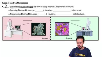
Types of Electron Microscopes
Location and Arrangement in Body Organs
The location and arrangement of these muscle types are crucial for their specific functions. Skeletal muscles are attached to bones and facilitate voluntary movements. Cardiac muscle is found exclusively in the heart, where its unique arrangement supports rhythmic contractions. Smooth muscle is located in the walls of hollow organs, such as the intestines and blood vessels, enabling involuntary movements like peristalsis and vasoconstriction.
Recommended video:
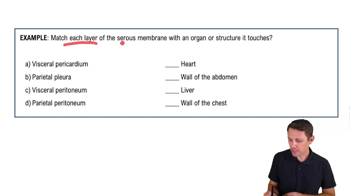
Organization of the Body: Serous Membrane Locations Example 1
Function of Muscle Types
Each muscle type serves distinct functions that are vital for bodily operations. Skeletal muscle is responsible for voluntary movements and posture, allowing for precise control. Cardiac muscle's function is to pump blood throughout the body, maintaining circulation. Smooth muscle controls involuntary actions, such as digestion and blood flow regulation, contributing to homeostasis and organ function.
Recommended video: