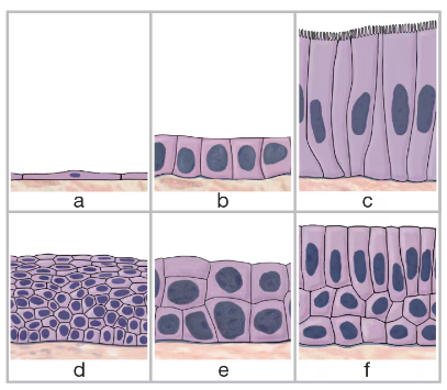
Identify the six types of epithelial tissue shown in the drawing below. d. ___
Verified step by step guidance
1
Step 1: Identify the simple squamous epithelium (a) by noting its single layer of flat, thin cells that allow for easy diffusion and filtration.
Step 2: Recognize the simple cuboidal epithelium (b) by its single layer of cube-shaped cells, often involved in secretion and absorption.
Step 3: Identify the simple columnar epithelium (c) by its single layer of tall, column-like cells, which may have microvilli or cilia for absorption or movement of substances.
Step 4: Observe the stratified squamous epithelium (d) characterized by multiple layers of cells with flat cells at the surface, providing protection against abrasion.
Step 5: Identify the stratified cuboidal epithelium (e) by its multiple layers of cube-shaped cells, typically found in ducts of glands, and the pseudostratified columnar epithelium (f) by its single layer of cells of varying heights that appear stratified but all touch the basement membrane, often with cilia.
Verified video answer for a similar problem:
This video solution was recommended by our tutors as helpful for the problem above
Video duration:
5m
Play a video:
0 Comments
Key Concepts
Here are the essential concepts you must grasp in order to answer the question correctly.
Types of Epithelial Tissue
Epithelial tissues are classified based on the number of cell layers and the shape of the cells. The main types include simple (one layer) and stratified (multiple layers), with shapes such as squamous (flat), cuboidal (cube-shaped), and columnar (tall and column-like). Understanding these types helps identify tissue functions and locations.
Simple epithelium consists of a single cell layer, facilitating processes like absorption and filtration. Stratified epithelium has multiple layers, providing protection against abrasion and pathogens. Recognizing the difference is crucial for identifying tissue roles in the body.
Some epithelial cells have specialized structures like cilia or microvilli to aid in movement or absorption. For example, ciliated columnar epithelium helps move mucus, while microvilli increase surface area for absorption. These features are key to understanding tissue function beyond basic classification.