Pearson+ LogoPearson+ Logo
Back

Sliding Filament Theory and the Sacromere quiz #3

Control buttons has been changed to "navigation" mode.
1/19
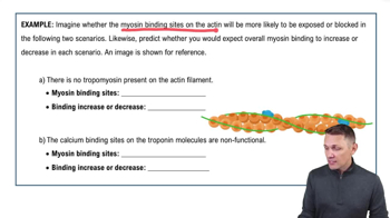
  • What roles do tropomyosin and troponin play in regulating muscle contraction?

    Tropomyosin blocks myosin binding sites on actin, preventing contraction. When calcium binds to troponin, troponin changes shape and moves tropomyosin away from the binding sites, allowing myosin to bind to actin and initiate contraction.
  • What is the function of the protein titin in the sarcomere?

    Titin is an elastic protein that anchors to the Z-disc and interacts with myosin, helping the sarcomere return to its original size after contraction or stretching. It is the largest known protein.
  • What is the sliding filament theory of muscle contraction?

    The sliding filament theory states that muscle contraction occurs when myosin (thick filament) pulls on actin (thin filament), causing the sarcomere to shorten as the filaments slide past each other without changing size.
  • How do myosin and actin interact during muscle contraction?

    Myosin heads reach out and pull on actin filaments, increasing the overlap between them and shortening the sarcomere.
  • What is the role of tropomyosin in muscle contraction?

    Tropomyosin is a thread-like protein that wraps around actin and blocks the myosin binding sites, preventing contraction when the muscle is relaxed.
  • How does troponin regulate muscle contraction?

    Troponin binds to calcium, which causes it to change shape and move tropomyosin away from the myosin binding sites on actin, allowing contraction to occur.
  • What is the function of titin in the sarcomere?

    Titin is an elastic protein that anchors to the Z-disc and interacts with myosin, helping the sarcomere return to its original size after contraction or stretching.
  • What structural feature marks the ends of a sarcomere and what does it anchor?

    The Z-disc marks the ends of a sarcomere and anchors the actin (thin) filaments.
  • What happens to the H-zone during muscle contraction?

    The H-zone, which is the region with only myosin, becomes smaller as actin slides over myosin and the overlap increases.
  • What are the I-band and A-band in a sarcomere, and what do they contain?

    The I-band is the light band containing only actin, while the A-band is the dark band containing both actin and myosin where they overlap.
  • What causes the striated appearance of skeletal muscle fibers?

    The striated appearance of skeletal muscle fibers is caused by the regular arrangement of thick (myosin) and thin (actin) filaments within the sarcomeres, creating alternating dark (A bands) and light (I bands) regions.
  • What is the functional unit of muscle contraction?

    The functional unit of muscle contraction is the sarcomere.
  • What is the contractile unit of a muscle?

    The contractile unit of a muscle is the sarcomere.
  • Why are skeletal muscle cells striated?

    Skeletal muscle cells are striated because of the organized, repeating pattern of sarcomeres, which contain alternating A bands (dark, with both actin and myosin) and I bands (light, with only actin).
  • What is the contractile unit of the muscle?

    The contractile unit of the muscle is the sarcomere.
  • What shortens during muscle contraction?

    During muscle contraction, the sarcomere shortens as the actin and myosin filaments slide past each other, increasing their overlap.
  • Which bands shorten during muscle contraction?

    During muscle contraction, the I band and the H zone shorten, while the A band remains the same length.
  • What is the basic unit of a muscle?

    The basic unit of a muscle is the sarcomere.
  • What are the thin myofilaments of muscle fiber composed of?

    The thin myofilaments of muscle fiber are composed of actin, along with regulatory proteins tropomyosin and troponin.