 Back
BackAnatomy & Physiology: Blood, Heart, and Blood Vessels
 2:36
2:36
Terms in this set (30)
Transport: oxygen, nutrients, wastes, hormones.
Regulation: body temperature, pH, fluid balance.
Protection: prevents blood loss and infection.
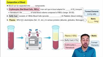
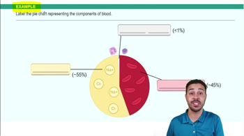
Blood consists of plasma (90% water, proteins, solutes) and formed elements (erythrocytes, leukocytes, platelets).
Because it consists of living cells (formed elements) suspended in a nonliving fluid matrix (plasma).
Biconcave, anucleate cells packed with hemoglobin; transport oxygen and carbon dioxide.
Protein with four globin chains, each bound to a heme group containing iron that binds oxygen.
Controlled by erythropoietin (EPO) from kidneys in response to hypoxia; requires iron, vitamin B12, folic acid.
Blood loss, decreased RBC production (iron deficiency, pernicious anemia), or increased RBC destruction (sickle-cell anemia).
Complete cells critical for defense; can leave bloodstream (diapedesis) and follow chemical signals (chemotaxis).
Granulocytes have visible granules (neutrophils, eosinophils, basophils); agranulocytes lack granules (lymphocytes, monocytes).
Cell fragments that form platelet plugs and release chemicals to promote clotting.
Vascular spasm, platelet plug formation, and coagulation (clotting).
Intrinsic: triggered by factors within blood; extrinsic: triggered by tissue factor from damaged tissue.
Based on presence of A and/or B antigens on RBCs; antibodies in plasma are opposite type.
Rh- mothers exposed to Rh+ fetal blood produce antibodies causing hemolysis in subsequent Rh+ pregnancies.
Located in mediastinum, size of a fist, two-thirds left of midline, apex points to left hip.
Epicardium (outer), myocardium (muscle), endocardium (inner lining).
Right atrium and ventricle pump deoxygenated blood to lungs; left atrium and ventricle pump oxygenated blood to body.
Prevent backflow; AV valves between atria and ventricles; semilunar valves at exits of ventricles.
Sinoatrial node, atrioventricular node, AV bundle, bundle branches, and Purkinje fibers coordinate heartbeat.
Branched cells with intercalated discs, self-excitable, longer refractory period, rely on aerobic respiration.
Ventricular filling (diastole), atrial contraction, ventricular systole, isovolumetric relaxation.
Cardiac output = stroke volume × heart rate.
Tunica intima (endothelium), tunica media (smooth muscle), tunica externa (connective tissue).
Elastic arteries (conducting), muscular arteries (distributing), arterioles (resistance vessels).
Continuous (least permeable), fenestrated (more permeable), sinusoid (most permeable).
Thinner walls, larger lumens, contain valves to prevent backflow, act as blood reservoirs.
Cardiac output, peripheral resistance, and blood volume.
MAP = diastolic pressure + 1/3 (systolic pressure - diastolic pressure).
Baroreceptors and chemoreceptors regulate cardiac output and vessel diameter via medullary centers.
Epinephrine, norepinephrine, angiotensin II increase pressure; atrial natriuretic peptide decreases pressure.