 Back
BackAnatomy & Physiology Core Concepts
 3:06
3:06

Terms in this set (30)
Anatomy is the study of body structure. Physiology is the study of body function and how the parts work together.
Gross anatomy studies structures visible to the naked eye. Forms include surface anatomy, regional anatomy, systemic anatomy, and clinical anatomy.
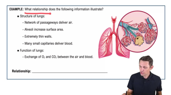
Microscopic anatomy studies structures too small to see without a microscope. Cytology is the study of cells. Histology is the study of tissues.
Levels include chemical, cellular, tissue, organ, organ system, and organism.
Examples: Integumentary (protection), skeletal (support), muscular (movement), nervous (control), cardiovascular (transport), respiratory (gas exchange), digestive (nutrient absorption), urinary (waste removal), endocrine (hormone regulation), lymphatic (immunity), reproductive (reproduction).
Homeostasis maintains stable internal conditions. Negative feedback reverses changes; positive feedback amplifies changes. Regulatory mechanisms include receptors, control centers, and effectors.
Superficial anatomy studies external body features and landmarks visible on the surface.
Body stands upright, feet forward, arms at sides, palms forward. Landmarks include head, neck, thorax, abdomen, limbs, etc.
Right upper, left upper, right lower, left lower quadrants, used to locate organs clinically.
Regions: right hypochondriac, epigastric, left hypochondriac, right lumbar, umbilical, left lumbar, right iliac, hypogastric, left iliac. Each contains specific organs like liver, stomach, intestines.
Terms include superior (above), inferior (below), anterior (front), posterior (back), medial (toward midline), lateral (away from midline), proximal (near trunk), distal (far from trunk).
Sagittal plane divides left and right, frontal (coronal) plane divides front and back, transverse plane divides top and bottom.
Major cavities: dorsal (cranial, spinal), ventral (thoracic, abdominopelvic). Serous membranes line cavities and reduce friction.
Epithelial (covering), connective (support), muscle (movement), nervous (control).
Cells tightly packed, polarity, avascular, high regeneration. Functions: protection, absorption, secretion. Specializations include cilia and microvilli.
Includes loose, dense, cartilage, bone, blood. Functions: support, protection, transport, energy storage.
Bone is rigid with mineralized matrix; supports and protects. Cartilage is flexible with chondrocytes; cushions joints. Types: hyaline, elastic, fibrocartilage.
Mucous, serous, cutaneous, and synovial membranes combine epithelial and connective tissues for protection and lubrication.
Superficial fascia stores fat, deep fascia surrounds muscles, subserous fascia lies under serous membranes, providing structural framework.
Skeletal muscle voluntary movement, striated; cardiac muscle involuntary, striated, heart; smooth muscle involuntary, non-striated, walls of organs.
Neurons transmit signals; neuroglia support and protect neurons in nervous tissue.
Swelling, redness, pain, heat due to increased blood flow and immune response. Regeneration repairs tissue damage.
Aging reduces epithelial renewal, bone density, muscle mass, and neural function. Cancer risk increases due to environmental factors and accumulated damage.
Protects body, regulates temperature, sensation. Layers: epidermis, dermis, hypodermis. Accessory structures include hair, nails, glands.
Skin color influenced by melanin, carotene, and blood flow. Melanin protects against UV damage; melanocytes increase melanin with sunlight exposure.
Sunlight converts skin cholesterol to vitamin D3, then to calcitriol in kidneys, aiding calcium absorption. Deficiency causes rickets.
Contains connective tissue, blood vessels, nerves, and sensory receptors. Cleavage lines indicate collagen fiber orientation important for surgery.
Eccrine sweat glands regulate temperature; apocrine glands produce scent; sebaceous glands secrete oil; mammary and ceruminous glands have specialized functions.
Nails are keratinized plates protecting fingertips. Formed by nail matrix cells dividing and hardening.
Includes inflammation, proliferation, and remodeling phases. Scar tissue forms from collagen deposition during healing.