Skip to main content
Anatomy & Physiology
My Course
Learn
Exam Prep
AI Tutor
Study Guides
Textbook Solutions
Flashcards
Explore
Try the app
My Course
Learn
Exam Prep
AI Tutor
Study Guides
Textbook Solutions
Flashcards
Explore
Try the app
Back
Anatomy & Physiology: Muscle and Nervous System Study Guide
You can tap to flip the card.
Connective tissue layers of muscle from outside to inside
You can tap to flip the card.
👆
Connective tissue layers of muscle from outside to inside
Epimysium
surrounds entire muscle,
Perimysium
surrounds fascicles,
Endomysium
surrounds individual muscle fibers.
Track progress
Control buttons has been changed to "navigation" mode.
1/30
Recommended videos
Guided course
8:11
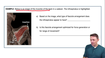
Fascicle Arrangements
6949
views
140
rank
Guided course
2:29
Fascicle Arrangements Example 1
4872
views
77
rank
Terms in this set (30)
Hide definitions
Connective tissue layers of muscle from outside to inside
Epimysium
surrounds entire muscle,
Perimysium
surrounds fascicles,
Endomysium
surrounds individual muscle fibers.
Types of fascicle arrangements
Parallel
: fibers run parallel (e.g., sartorius),
Pennate
: feather-like (uni-, bi-, multipennate),
Convergent
: broad origin to narrow insertion,
Circular
: surrounds openings (e.g., orbicularis oris).
Order of skeletal muscle organization from largest to smallest
Muscle → Fascicle → Muscle fiber (cell) → Myofibril → Sarcomere → Myofilaments.
Sarcomere regions and their filament composition
Z disc
: sarcomere boundary,
A band
: thick filaments,
I band
: thin filaments only,
H zone
: thick filaments only,
M line
: center of sarcomere.
What happens to sarcomere bands during muscle contraction?
I band
shortens,
H zone
shortens,
A band
stays the same length.
Main proteins in thick and thin filaments
Thick filament:
Myosin
. Thin filament:
Actin
,
Troponin
,
Tropomyosin
.
Role of tropomyosin and troponin in muscle contraction
Tropomyosin
blocks actin binding sites;
Troponin
binds calcium and moves tropomyosin to expose active sites.
Sequence of events in the sliding filament theory
Calcium released → binds troponin → tropomyosin moves → myosin binds actin → power stroke → ATP binds myosin → myosin detaches → ATP hydrolyzed → head resets.
Why is ATP needed in muscle contraction?
ATP is required for
myosin detachment
from actin,
re-cocking
the myosin head, and powering the
calcium pump
.
Role of calcium in muscle contraction
Calcium binds to troponin, causing tropomyosin to move and expose active sites on actin, enabling contraction.
Steps at the neuromuscular junction to initiate muscle contraction
Action potential arrives → calcium enters neuron → acetylcholine released → ACh binds receptors → sodium channels open → depolarization → muscle action potential → calcium released from SR → contraction.
Function of acetylcholine and acetylcholinesterase at NMJ
Acetylcholine stimulates muscle contraction; acetylcholinesterase breaks down ACh to stop stimulation.
Difference between isometric and isotonic muscle contractions
Isometric
: tension changes, length stays same (e.g., holding dumbbell still).
Isotonic
: muscle length changes; concentric shortens, eccentric lengthens.
Components of a lever system in the body
Fulcrum
: pivot point,
Effort
: force applied,
Load
: resistance.
Functions of the masseter and sternocleidomastoid muscles
Masseter: chewing. Sternocleidomastoid: rotates and flexes neck.
Muscles involved in breathing
Diaphragm and intercostals.
Four rotator cuff muscles (SITS)
Supraspinatus, Infraspinatus, Teres minor, Subscapularis.
Muscle adaptation: hypertrophy
Increase in muscle size due to increased workload, resistance training, and increased myofibrils/protein synthesis.
Neuron parts and their functions
Dendrites: receive signals, Cell body: control center, Axon: conducts impulse, Synaptic terminal: releases neurotransmitter.
Myelin-producing cells in CNS and PNS
CNS: oligodendrocytes. PNS: Schwann cells.
Resting membrane potential and ion distribution
About -70 mV; more sodium outside, more potassium inside the cell.
What occurs during depolarization and hyperpolarization?
Depolarization: membrane becomes less negative (sodium enters). Hyperpolarization: membrane becomes more negative (potassium leaves or chloride enters).
Threshold potential for triggering an action potential
Usually around -55 mV.
Saltatory conduction
Impulse jumps from node to node along myelinated axons, increasing conduction speed.
Difference between absolute and relative refractory periods
Absolute: neuron cannot fire again. Relative: neuron can fire if stimulus is strong enough.
Inhibitory neurotransmitter example
GABA
is an inhibitory neurotransmitter.
Functions of the frontal, temporal, parietal, and occipital lobes
Frontal: motor, personality, planning. Temporal: hearing, memory. Parietal: somatosensory. Occipital: vision.
Primary motor and somatosensory cortex functions
Primary motor cortex controls voluntary movement; primary somatosensory cortex processes touch sensation.
Functions of the hypothalamus
Regulates temperature, hunger/thirst, endocrine control, and autonomic nervous system.
Role of the cerebellum
Coordinates movement, balance, and motor learning.
 BackBack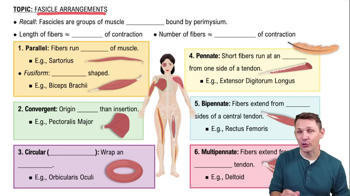
 8:11
8:11