 Back
BackAnatomy & Physiology: Musculoskeletal Structures and Muscles of Upper and Lower Extremities
 5:30
5:30
Terms in this set (22)
A gliding synovial joint between the acromion of the scapula and the clavicle.
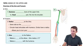
A ball and socket synovial joint allowing triaxial movement between the humerus and scapula.
A uniaxial hinge synovial joint allowing flexion and extension between humerus, radius, and ulna.
A uniaxial pivot synovial joint allowing pronation and supination of the forearm.
Abduction of the shoulder; anterior head does flexion and medial rotation, posterior head does extension and lateral rotation.
Originates on the medial clavicle and sternum; actions include flexion, adduction, and medial rotation of the humerus.
Originates on thoracic and lumbar vertebrae, sacrum, and iliac crest; actions include extension, adduction, and medial rotation of the humerus.
Infraspinatus and teres minor muscles laterally rotate the humerus at the shoulder joint.
Flexes the shoulder and elbow; inserts on the radial tuberosity.
Extends the shoulder and elbow; inserts on the olecranon process of the ulna.
Brachialis and brachioradialis flex the elbow; pronator teres pronates the forearm.
Flexor carpi radialis causes flexion and abduction of the wrist; flexor carpi ulnaris causes flexion and adduction.
Both muscles cause extension and abduction of the wrist.
Includes ilium, ischium, and pubis bones forming the coxal bone with features like iliac crest, ischial tuberosity, and pubic symphysis.
A ball and socket synovial joint between the femur head and acetabulum of the pelvis.
Gluteus medius and gluteus minimus abduct the hip.
Extends the knee; rectus femoris also flexes the hip.
Extend the hip and flex the knee; includes biceps femoris, semimembranosus, and semitendinosus.
Causes dorsiflexion of the ankle.
Flexes the knee and causes plantar flexion of the ankle; inserts via Achilles tendon on the calcaneus.
Includes medial collateral ligament (MCL), lateral collateral ligament (LCL), anterior cruciate ligament (ACL), and posterior cruciate ligament (PCL).
Tensor fascia latae (TFL) originates on the iliac crest and inserts on the IT band to stabilize the knee.