The lens in a microscope that is closest to the eye, used to view the magnified image.
Objective Lens
The primary lenses on a microscope that magnify the specimen; commonly color-coded as red, yellow, and blue for different magnifications.
Mechanical Stage Controls
Knobs on a microscope used to move the slide precisely on the stage for better viewing.
Iris Diaphragm
A component of the microscope that adjusts the amount of light reaching the specimen.
Anatomical Landmarks
Specific points on the body used as reference for anatomical study and description.
Three Major Body Cavities
The dorsal cavity, ventral cavity, and abdominopelvic cavity that house major organs.
Nine Abdominal Pelvic Regions
Divisions of the abdomen used to locate organs: right and left hypochondriac, epigastric, right and left lumbar, umbilical, right and left iliac, and hypogastric regions.
Directional Terminology
Terms used to describe locations on the body, such as anterior, posterior, medial, lateral, proximal, and distal.
Planes of the Body
Imaginary lines used to divide the body: midsagittal, parasagittal, transverse, coronal/frontal, and oblique.
Interphase
The cell cycle phase where the cell grows and DNA is replicated before mitosis.
Prophase
The first stage of mitosis where chromatin condenses into chromosomes and the nuclear envelope breaks down.
Metaphase
The mitosis phase where chromosomes align at the cell's equatorial plate.
Anaphase
The mitosis phase where sister chromatids separate and move toward opposite poles.
Telophase and Cytokinesis
Final mitosis stages where nuclear envelopes reform and the cell divides into two daughter cells.
Simple Squamous Epithelium
A single layer of flat cells that allows for diffusion and filtration.
Simple Cuboidal Epithelium
A single layer of cube-shaped cells involved in secretion and absorption.
Simple Columnar Epithelium
A single layer of tall cells that often have microvilli and are involved in absorption and secretion.
Pseudostratified Columnar Epithelium
A single layer of cells with varying heights that appears stratified; often ciliated for moving mucus.
Stratified Squamous Epithelium
Multiple layers of flat cells that protect underlying tissues in areas subject to abrasion.
Loose Connective Tissue: Areolar
A connective tissue with a loose arrangement of fibers that cushions and protects organs.
Dense Regular Connective Tissue
Tightly packed collagen fibers arranged in parallel, providing strong attachment in tendons and ligaments.
Hyaline Cartilage
A translucent cartilage that provides support with some flexibility; found in the nose and trachea.
Skeletal Muscle
Voluntary muscle tissue characterized by striations and multiple nuclei per cell.
Cardiac Muscle
Involuntary, striated muscle found only in the heart with intercalated discs for synchronized contraction.
Smooth Muscle
Involuntary muscle tissue without striations found in walls of hollow organs.
Nervous Tissue
Tissue composed of neurons and supporting cells that transmit electrical signals throughout the body.
 Back
Back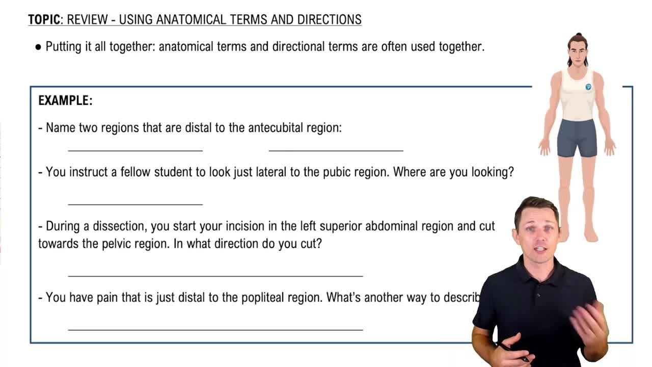
 03:47
03:47