 Back
BackAnatomy & Physiology: Tissues and Integumentary System

 03:32
03:32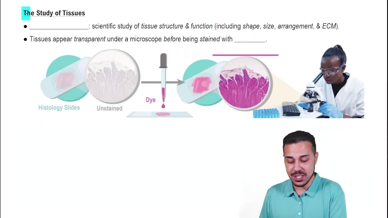
Terms in this set (50)
Cell, tissue, organ, organ system
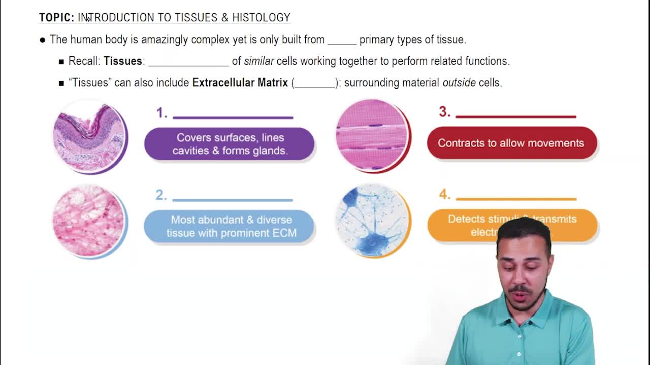
Epithelial tissue
Complementarity of structure and function
They lack blood vessels but contain nerve fibers.
Basement membrane
Apical cells are the most exposed and visible layer, while basal cells are constantly dividing and vary in shape.
Simple squamous epithelium
Found in kidney tubules and secretory ducts of small glands.
Simple columnar epithelium
All cells rest on the basement membrane but nuclei are at different heights, creating the illusion of multiple layers.
Transitional epithelium
Endocrine glands secrete hormones directly into blood or interstitial fluid, exocrine glands secrete onto epithelial surfaces via ducts.
Goblet cell
Simple alveolar (acinar) gland
Apocrine secretion
Holocrine secretion
Extracellular matrix composed of ground substance and fibers
It is found in every organ system and functions to bind, support, protect, insulate, and transport.
Collagen fibers
Mast cells
Areolar connective tissue
Adipose tissue consists mostly of fat-storing cells with nuclei pushed to the side by large fat droplets.
Reticular connective tissue
Closely packed, parallel collagen fibers aligned with the direction of pull.
Its fibers run in multiple directions, resisting tension from many angles.
Cartilage is avascular and relies on slow diffusion for nutrients.
Chondroblasts secrete new cartilage matrix, chondrocytes are mature cells maintaining the matrix.
Hyaline cartilage
Fibrocartilage
Inorganic calcium salts deposited around collagen fibers
It develops from mesenchyme and consists of living cells suspended in a non-living fluid matrix (plasma).
Skeletal muscle tissue
Cardiac muscle cells are branched, uninucleated, and connected by intercalated discs.
Neurons generate and conduct impulses; neuroglia support and protect neurons.
Serous membranes; they are wet membranes.
Inflammation causes blood vessels to dilate and become permeable for immune cells and clotting proteins.
Cardiac muscle and nervous tissue in brain/spinal cord
It contains multiple tissue types working together to perform complex functions.
Stratum basale, stratum spinosum, stratum granulosum, stratum lucidum, stratum corneum
Stratum basale
Dendritic (Langerhans) cells
Melanin forms a pigment shield over the nucleus blocking ultraviolet radiation.
They undergo apoptosis and accumulate keratin as they move away from blood supply.
Papillary layer (areolar connective tissue) and reticular layer (dense irregular connective tissue)
Anchors skin, stores fat, cushions blows, and insulates against heat loss.
Arrector pili muscle
Sebaceous glands producing sebum
Eccrine glands function in thermoregulation and open directly onto skin; apocrine glands open into hair follicles and activate at puberty.
Melanoma
Second-degree burn