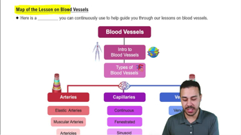
What are the three main types of blood vessels and their primary functions?
Arteries carry blood away from the heart (usually oxygenated), capillaries serve cellular needs by direct exchange, and veins carry blood toward the heart (usually deoxygenated).
What are the three layers (tunics) of most blood vessel walls?
Tunica intima (innermost), tunica media (middle, smooth muscle and elastin), and tunica externa (outer collagen fibers).
What is the structure and function of the tunica intima?
Innermost layer lined by endothelium (simple squamous epithelium) that reduces friction; includes a subendothelial connective tissue layer in vessels larger than 1 mm.
What composes the tunica media and what is its role?
Made mostly of smooth muscle and elastin sheets; controls vessel diameter via vasoconstriction and vasodilation, regulating blood flow and pressure.
Describe the tunica externa and its special features.
Outermost layer of loose collagen fibers that protect and anchor vessels; contains nerve fibers, lymphatic vessels, and in large vessels, the vasa vasorum to nourish the outer wall.
How do arteries and veins differ in tunica media and tunica externa thickness?
Arteries have a thicker tunica media than tunica externa; veins have a thicker tunica externa than tunica media.
What are elastic arteries and their function?
Thick-walled arteries with large lumens near the heart; act as pressure reservoirs that expand and recoil to maintain continuous blood flow.
What distinguishes muscular arteries from elastic arteries?
Muscular arteries have thicker tunica media with more smooth muscle and less elastin; they distribute blood to organs and actively regulate blood flow.
What are arterioles and their role in blood flow?
Smallest arteries controlling blood flow into capillary beds via vasodilation and vasoconstriction; also called resistance arteries due to their role in blood flow resistance.
What is the structure of capillaries and their main function?
Microscopic vessels with walls of only tunica intima (endothelium and basal lamina); function in exchange of gases, nutrients, wastes, and hormones between blood and tissues.
Name the three types of capillaries and a key feature of each.
Continuous capillaries: least permeable, most common; fenestrated capillaries: have pores for increased permeability; sinusoidal capillaries: most permeable with large gaps, found in liver, spleen, bone marrow.
What are pericytes and their function in capillaries?
Spider-shaped stem cells that stabilize capillary walls, control permeability, and assist in vessel repair.
What is a capillary bed and what controls blood flow through it?
An interwoven network of capillaries between arterioles and venules; blood flow is regulated by diameter of terminal arterioles and precapillary sphincters responding to local chemical signals.
What is a vascular shunt in capillary beds?
A channel (metarteriole and thoroughfare channel) that directly connects an arteriole to a venule, bypassing true capillaries.
What are precapillary sphincters and their role?
Rings of smooth muscle around true capillaries that regulate blood flow into capillary beds based on local chemical conditions.
How do veins differ structurally from arteries?
Veins have thinner walls, larger lumens, thinner tunica media, and thicker tunica externa; they contain valves to prevent backflow and act as blood reservoirs.
What adaptations help veins return blood to the heart despite low pressure?
Large lumens reduce resistance, venous valves prevent backflow, and venous sinuses provide thin-walled channels for blood.
What causes varicose veins and what factors contribute?
Dilated, painful veins due to incompetent valves; contributed by heredity, prolonged standing, obesity, pregnancy, and increased venous pressure.
What are vascular anastomoses and their significance?
Interconnections of blood vessels providing alternate pathways for blood flow, ensuring continuous circulation if one vessel is blocked.
Where are arterial anastomoses commonly found and where are they absent?
Common in joints, abdominal organs, brain, and heart; absent in retina, kidneys, and spleen.
 Back
Back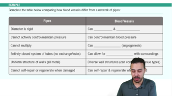
 3:38
3:38