 Back
BackCh 10 Quiz Power Point A+P I
 6:31
6:31
Terms in this set (58)
What is all muscle tissues one basic function?
To generate force (muscle tension)
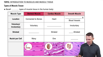
What are 3 types of muscle tissue in the body?
Cardiac Muscle Tissue
Smooth Muscle Tissue
Skeletal Muscle Tissue
What are the 5 properties of cardiac muscles?
Located in cardiac tissue
Striated in appearance
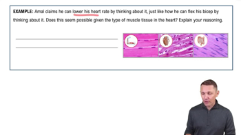
Involuntary Control
Single central nucleus
Intercalated discs with gap junctions/desmosomes
Cardiac Muscle tissue has more _______ and ________.
Sarcoplasm
Mitochondria
Which muscle has more calcium allowing it to contract 10-15 times longer than skeletal tissue
Cardiac Muscle Tissue
Involuntary built-in rhythm which functions as a pacemaker to initiate the cardiac cycle
Autothythmicity
What are the 4 properties of smooth muscle tissue?
Not Striated
Involuntary
(Visceral) Lines hallow organs and blood vessels
(Multiunit) Individual fibers
What muscle tissue does the Calcium move slowly out of the cell (longer presence of calcium in the cell)?
Smooth Muscle Tissue
Smooth muscle fibers can stretch considerably without developing tension (can change pressure)
Useful for maintaining blood pressure
Stress-Relaxation Response
What are the 5 properties of skeletal muscle tissue?
attaches to bone, skin, or fascia
striated
Multinucleated cells
contractions are voluntary
fibers are formed by fusion of many embryonic myoblasts giving each fiber multiple nuclei
What causes skeletal muscle tissue to be striated?
due to alternating light and dark bands of protein within the cells
What is the purpose of skeletal muscle tissue? (5 things)
Produce body movements
Stabilizes body positions
Regulates organ volumes
Moves substances
Produces heat
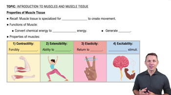
Both muscle & nerve tissues produce action potentials
Excitability
Ability to shorten and generate force
Contractility
Ability to be stretched without damaging the tissue
Extensibility
Ability to return to original shape after being stretched
Elasticity
extends from tendon and surrounds entire muscle
Epimysium
Deep to epimysium; surrounds bundles (fascicles) of 10-100 muscle cell/fibers
Perimysium
Separates individual muscle cells
Endomysium
Plasma membrane surrounding muscle cells
Sarcolemma
cytoplasm of muscle cell
Sarcoplasm
Similar to ER
Sarcoplasmic Reticulum (SR)
Contains many mitochondria for ATP production
Also contains myoglobin and myofibrils
Sarcoplasm
Forms web-like network surrounding myofibrils
Houses Calcium
Sarcoplasmic Reticulum (SR)
A protein within the sarcoplasm that stores O2 for ATP production and aerobic respiration
Gives muscles their red color
Myoglobin
100s-1000s of tiny threads that run parallel through muscle fibers
Made up of bundles of specialized proteins; allow for contraction (contractile proteins)
Myofibrils
Contractile elements of skeletal muscle
Made up of thick and thin filaments
Myofibrils
Thick and thin filaments are called
myofilaments
What are the 3 types of proteins that can be found in myofibrils
Contractile Proteins
Regulatory Proteins
Structural Proteins
Which protein generates tension?
Contractile Proteins
Which protein dictates when a fiber may contract?
Regulatory Proteins
Which protein maintains proper myofilament alignment and fiber stability?
Structural Proteins
What are the 3 types of myofilaments called?
Thick Filaments
Thin Filaments
Elastic Filaments
What type of myofilament is contractile protein?
Thick Filaments
What type of myofilament is structural protein?
Elastic Filament
What is thick filament made up of?
Myosin
What is the thin filament made up of?
Actin (contractile), Tropomyosin and troponin (regulatory)
What is Elastic filament made up of?
Titin (stabilizes myofibril structure)
Dystrophin (resists excessive stretching)
Sarcomeres are separated by _____
Z discs
Zigzags of dense protein
Z discs
What is the compartment between Z discs called?
Sarcomeres
Deep inward extensions of sarcolemma; surround each myofibril
Transverse Tubules (T tubules)
What is the function of Transverse Tubules?
Carry muscle APs down into cell
Stores Calcium in a relaxed muscle
release of calcium triggers muscle contrations
Sarcoplasmic Reticulum (SR)
a motor protein helps convert ATP to a usable source of energy
Myosin
Each head of myosin has active site that binds with _____
Actin
Shaped like 2 twisted golf clubs heads and a tail
myosin
Made up of actin, troponin, tropomyosin
Thin Filaments
twist into a helix and contains myosin-binding sites where myosin heads can attach
actin
Strands cover myosin-binding sites in relaxed muscle
Tropomyosin
Small globular regulatory protein; holds tropomyosin in place; assists with turning contractions on and off
Troponin
large-molecule protein responsible for much of the elasticity and extensibility of myofibrils
Titin
Anchors thick filaments to the M line (center of sarcomere) and the Z disc
Titin
The swiveling of the myosin heads pulling the thin filaments toward the M line (the center of the sarcomere)
Sliding Filament Mechanism
the swiveling of myosin heads pulling the actin toward the M line (ATP hydrolyzed)
Power Stroke
area in which nerve and muscle meet
they do not touch
Neuromuscular Junction (NMJ)
Small space between where the nerve and muscle meet in Neuromuscular Junction (NMJ)
Synaptic Cleft
swelling of the axon terminals
Synaptic end bulbs