 Back
BackConnective Tissue, Muscle, and Neuron Histology - Anatomy & Physiology
 06:37
06:37
Terms in this set (23)
Parallel collagen fibers tightly packed with fibroblasts. Provides firm attachment, conducts muscle pull, reduces friction, and stabilizes bones. Found in tendons, aponeuroses, and ligaments.
Thick collagen fibers woven randomly. Provides structural strength and resists forces from many directions. Located in dermis, digestive tract submucosa, and fibrous capsules of joints and organs.
Cartilage with thick type 1 collagen fibers. Acts as shock absorber, cushions joints, and withstands pressure. Found in intervertebral discs, pubic symphysis, and knee menisci.
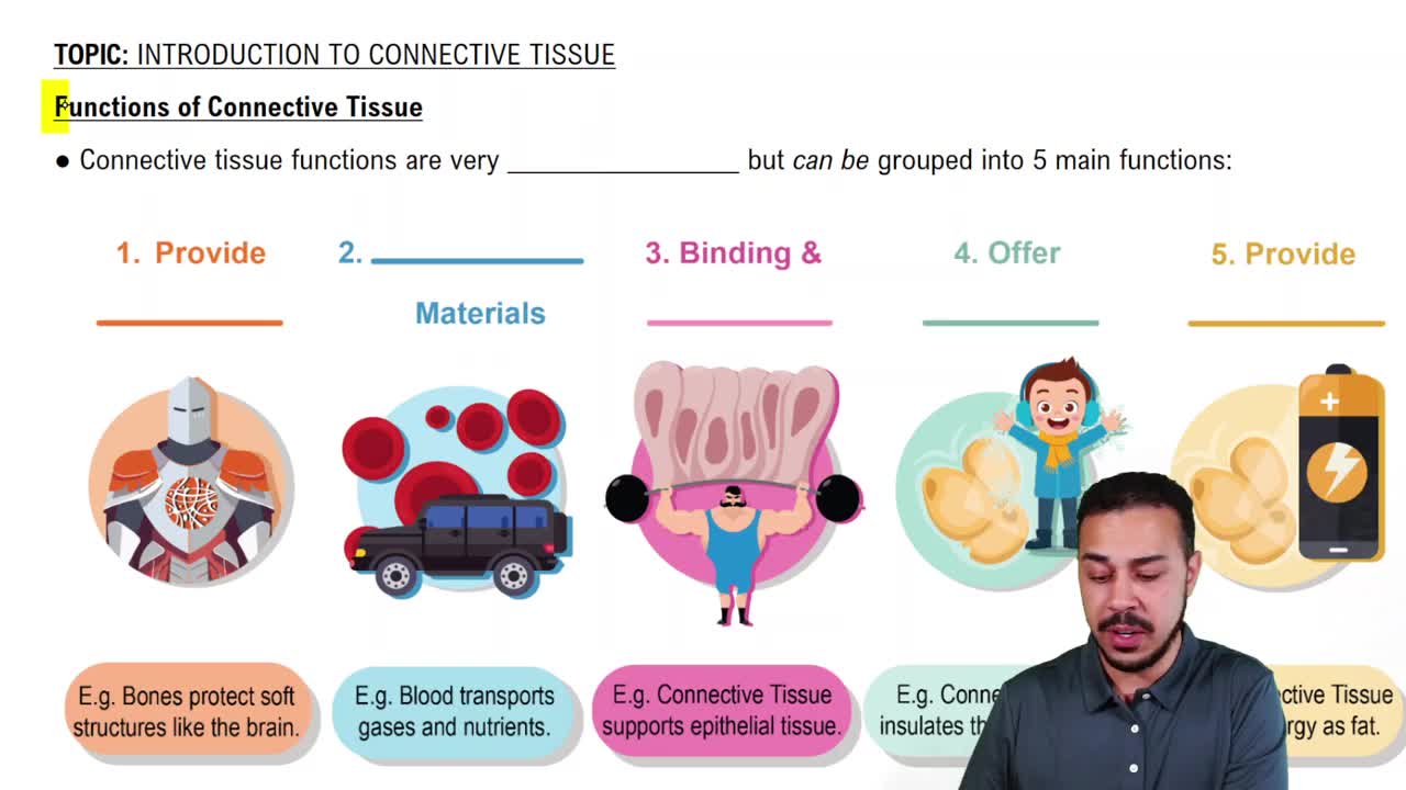
Dense, hard outer bone layer with osteons. Provides rigid support, protects organs, stores calcium, and acts as lever for muscles. Forms external layer and diaphysis of long bones.
Porous bone with trabeculae. Reduces skeleton weight, absorbs impact, and houses red marrow for blood cell production. Found inside short, flat, irregular bones and epiphyses of long bones.
Fat cells with large lipid droplets. Stores energy, insulates against heat loss, and cushions organs. Located subcutaneously, around kidneys, behind eyeballs, and in yellow bone marrow.
Loose, web-like matrix with collagen, elastic, and reticular fibers. Binds epithelia to deeper tissues, cushions organs, holds tissue fluid, and supports immune defense. Found beneath epithelial sheets and around vessels.
Fluid connective tissue with erythrocytes, leukocytes, and platelets in plasma. Transports oxygen, nutrients, wastes, regulates temperature, and supports immune defense. Circulates in cardiovascular system.
Glassy cartilage with fine collagen fibers in chondroitin sulfate matrix. Provides smooth joint surfaces, flexibility, and airway support. Found on long bone ends, costal cartilages, nose, larynx, trachea, and bronchi.
Delicate mesh of reticular fibers and cells forming soft internal skeleton. Supports free blood cells and immune system. Located in lymph nodes, spleen, bone marrow, and liver.
Cartilage rich in elastic fibers. Provides elasticity and resilience, allowing shape recovery. Found in external ear (pinna) and epiglottis.
Gel-like extracellular matrix component of connective tissue. Allows diffusion of nutrients and wastes, acts as barrier to pathogens, and varies from fluid to calcified depending on tissue.
These are loose connective tissues characterized by a loose fiber arrangement and diverse cell types.
Classified as dense connective tissue due to tightly packed collagen fibers arranged irregularly.
Classified as fluid connective tissue because it is a liquid matrix with suspended cells.
These are supporting connective tissues providing structural support and protection.
Thick, straight, unbranched bundles of collagen protein. Provide high tensile strength to resist pulling forces.
Thin, wavy, branched strands made of elastin and fibrillin. Provide elasticity allowing tissues to stretch and snap back.
Thin, short, delicate mesh networks of type III collagen. Form supportive framework holding cells in soft organs.
Striated, multinucleated muscle under voluntary control. Produces body movement, posture, heat, and voluntary sphincters. Found attached to skeleton and in tongue, pharynx, diaphragm.
Striated, branched muscle with intercalated discs. Contracts involuntarily to pump blood and maintain blood pressure. Located in myocardium of the heart.
Non-striated, spindle-shaped involuntary muscle. Propels substances via peristalsis, regulates blood vessel diameter, controls airflow, and organ functions. Found in walls of hollow organs and blood vessels.
Specialized nervous system cell that receives, processes, and transmits electrical and chemical signals. Coordinates sensory input, motor output, cognition, and homeostasis. Located in CNS and PNS.