Skip to main content
Anatomy & Physiology
My Course
Learn
Exam Prep
AI Tutor
Study Guides
Textbook Solutions
Flashcards
Explore
Try the app
My Course
Learn
Exam Prep
AI Tutor
Study Guides
Textbook Solutions
Flashcards
Explore
Try the app
Back
Set #6 Pelvis, Legs, and Lower Limbs Anatomy
You can tap to flip the card.
Metacarpals (Numbered I – V)
You can tap to flip the card.
👆
Metacarpals (Numbered I – V)
The 5 bones forming the palm of the hand, numbered starting with the thumb as Metacarpal I.
Track progress
Control buttons has been changed to "navigation" mode.
1/35
Recommended videos
Guided course
02:05
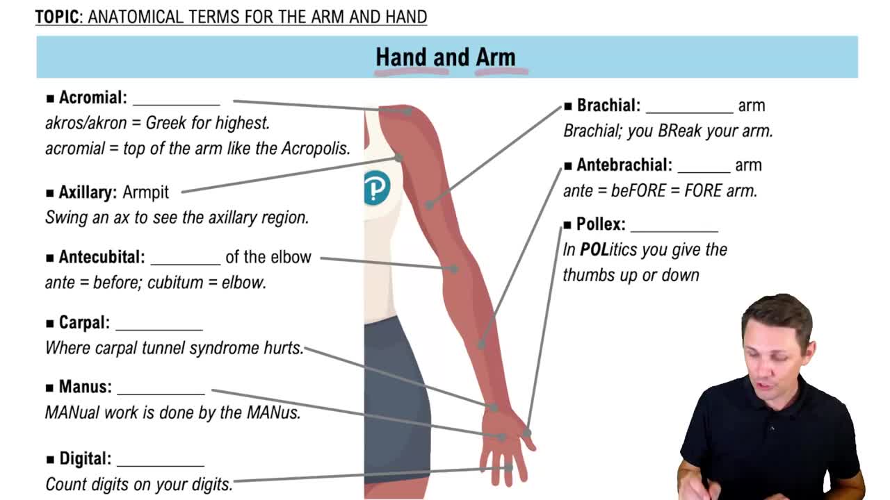
Anatomical Terms for the Arm and Hand Example 1
5474
views
128
rank
Guided course
03:17
The Arm and Hand
7905
views
153
rank
1
comments
Terms in this set (35)
Hide definitions
Metacarpals (Numbered I – V)
The 5 bones forming the palm of the hand, numbered starting with the thumb as Metacarpal I.
Phalanges (Hand & Foot)
Bones forming the fingers or toes. Fingers/toes II-V have 3 phalanges; the thumb/big toe has 2.
Proximal / Middle / Distal phalanges
Proximal: closest to palm/sole. Middle: middle bone (absent in thumb/big toe). Distal: tips of fingers/toes.
Pelvic girdle / Coxal bone
Hip bone formed by fusion of ilium, ischium, and pubis. Must differentiate right vs. left side.
Acetabular fossa
Deep cup-like depression on lateral coxal bone surface where all three regions fuse; receives the femur head.
Obturator foramen
Large closed opening formed by ischium and pubis bones allowing passage of blood vessels and nerves.
Ilium
Largest, superior, flared wing-like section of the coxal bone.
Iliac fossa
Smooth, shallow internal concave surface of the flared ilium wing.
Iliac crest
Thickened superior border ridge of the ilium; palpable as the 'hip bone' when hands rest on hips.
Anterior superior & Anterior inferior iliac spines
ASIS: bony projection at front tip of iliac crest. AIIS: bony bump directly below ASIS.
Posterior superior & Posterior inferior iliac spines
PSIS: bony projection at back tip of iliac crest. PIIS: bony bump directly below PSIS.
Auricular surface (of Ilium)
Rough ear-shaped internal patch on ilium that joins with sacrum to form sacroiliac joint.
Greater sciatic notch
Large, deep J-shaped indentation on posterior border of ilium below the PIIS.
Ischium
Inferior, posterior region of the coxal bone, known as the 'sit bone.'
Ischial tuberosity
Rough, thickened loop of bone on lowest ischium curve; bears weight when sitting.
Ischial spine
Sharp bony projection pointing medially just above the ischial tuberosity.
Ischial ramus
Flat bar of bone extending forward from tuberosity to merge with inferior pubis ramus.
Pubis
Anterior, inferior region of the coxal bone.
Superior & Inferior pubic rami
Superior ramus projects back toward acetabulum; inferior ramus projects down toward ischial ramus.
Pubic tubercle
Small forward bump on superior pubic ramus near the body's midline.
Pubic symphysis
Fibrocartilage pad joint uniting left and right pubic bones at the midline.
Femur
Thigh bone; longest, heaviest, and strongest bone in the human body.
Femoral head
Smooth spherical proximal ball pointing medially to articulate with acetabulum.
Femoral neck
Constricted bridge connecting femoral head to the main shaft.
Fovea capitis
Small pit in center of femoral head serving as attachment for an internal ligament.
Greater trochanter
Large rough projection pointing upward on lateral proximal femur shaft.
Lesser trochanter
Smaller projection pointing posteromedially on proximal femur shaft below the neck.
Linea aspera
Prominent vertical ridge on posterior femur shaft for muscle attachment.
Medial & Lateral condyles (Femur)
Large distal projections articulating with tibia; medial condyle is on same side as femoral head.
Medial & Lateral epicondyles (Femur)
Rough bumps located directly above respective condyles on distal femur.
Intercondylar fossa
Deep U-shaped groove separating medial and lateral condyles on posterior distal femur.
Patella
Kneecap; small triangular sesamoid bone embedded in quadriceps tendon.
Tibia
Shinbone; massive medial, weight-bearing bone of the lower leg.
Medial & Lateral condyles (Tibia)
Flattened smooth superior surfaces at proximal tibia that articulate with femur condyles.
Tibial tuberosity
Rough projection on anterior midline of tibia below condyles; attachment for patellar ligament.
 BackBack
 02:05
02:05