Join thousands of students who trust us to help them ace their exams!
Multiple Choice
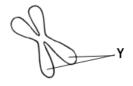
The structures labeled Y in the image below are called:
A
Centromeres.
B
Centrioles.
C
Sister chromatids.
D
Spindles.
0 Comments
Verified step by step guidance
1
Examine the image provided. The structure shown is a chromosome, which is typically X-shaped during certain stages of cell division.
Identify the labeled part 'Y' in the image. It points to the region where the two identical halves of the chromosome are joined.
Understand that each half of the chromosome is called a chromatid. When they are joined together, they are referred to as sister chromatids.
Recall that the centromere is the region where the sister chromatids are most tightly connected, but the label 'Y' is pointing to the entire structure of the joined chromatids.
Conclude that the structures labeled 'Y' are sister chromatids, as they represent the two identical copies of a chromosome connected together.


 Verified step by step guidance
