What is the main purpose of genome-wide association studies (GWAS)? How can information from GWAS be used to inform scientists and physicians about genetic diseases?
Table of contents
- 1. Introduction to Genetics51m
- 2. Mendel's Laws of Inheritance3h 37m
- 3. Extensions to Mendelian Inheritance2h 41m
- 4. Genetic Mapping and Linkage2h 28m
- 5. Genetics of Bacteria and Viruses1h 21m
- 6. Chromosomal Variation1h 48m
- 7. DNA and Chromosome Structure56m
- 8. DNA Replication1h 10m
- 9. Mitosis and Meiosis1h 34m
- 10. Transcription1h 0m
- 11. Translation58m
- 12. Gene Regulation in Prokaryotes1h 19m
- 13. Gene Regulation in Eukaryotes44m
- 14. Genetic Control of Development44m
- 15. Genomes and Genomics1h 50m
- 16. Transposable Elements47m
- 17. Mutation, Repair, and Recombination1h 6m
- 18. Molecular Genetic Tools19m
- 19. Cancer Genetics29m
- 20. Quantitative Genetics1h 26m
- 21. Population Genetics50m
- 22. Evolutionary Genetics29m
18. Molecular Genetic Tools
Methods for Analyzing DNA
Problem 15b
Textbook Question
The bacteriophage lambda genome can exist in either a linear form or a circular form.
Diagram the resulting fragments as they would appear on an agarose gel after electrophoresis.
 Verified step by step guidance
1
Understand the context: The bacteriophage lambda genome can exist in two forms—linear and circular. Linear DNA will produce distinct fragments when digested with restriction enzymes, while circular DNA may produce different patterns due to the absence of free ends.
Identify the restriction enzyme used: Determine which restriction enzyme is applied to the DNA. Each enzyme cuts at specific recognition sites, producing fragments of predictable sizes. For example, EcoRI recognizes the sequence GAATTC and cuts between G and A.
Predict the fragment sizes: For the linear form, calculate the sizes of fragments based on the known positions of restriction sites along the genome. Use the genome map to determine the distances between consecutive restriction sites.
Consider the circular form: In circular DNA, the restriction enzyme cuts at the same recognition sites, but the absence of free ends means the fragments may differ. For example, one fragment may represent the remainder of the circular genome after the first cut.
Diagram the gel: Represent the fragments as bands on an agarose gel. Larger fragments will migrate slower and appear closer to the top, while smaller fragments will migrate faster and appear closer to the bottom. Label the bands with their corresponding fragment sizes for both linear and circular forms.
 Verified video answer for a similar problem:
This video solution was recommended by our tutors as helpful for the problem above
Video duration:
3mPlay a video:
Was this helpful?
Key Concepts
Here are the essential concepts you must grasp in order to answer the question correctly.
Bacteriophage Lambda Genome Structure
The bacteriophage lambda genome can exist in two forms: linear and circular. The linear form is typically found during the lytic cycle, where the virus injects its DNA into a host cell, while the circular form is associated with the lysogenic cycle, where the viral DNA integrates into the host genome. Understanding these forms is crucial for predicting how the DNA will behave during gel electrophoresis.
Recommended video:
Guided course

Bacteriophage Life Cycle
Gel Electrophoresis
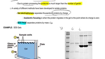
Gel electrophoresis is a technique used to separate DNA fragments based on their size. When an electric current is applied, negatively charged DNA moves towards the positive electrode, with smaller fragments migrating faster than larger ones. This method allows visualization of the DNA fragments, which is essential for analyzing the results of the bacteriophage lambda genome's different forms.
Recommended video:
Guided course

Proteomics
Fragmentation Patterns
The fragmentation patterns of DNA during gel electrophoresis depend on the form of the genome and the restriction enzymes used. Linear DNA typically produces distinct bands corresponding to the sizes of the fragments generated, while circular DNA may appear as a single band or multiple bands depending on its conformation and any cuts made. Recognizing these patterns is key to accurately diagramming the results on an agarose gel.
Recommended video:
Guided course

Segmentation Genes

 7:40m
7:40mWatch next
Master Methods for Analyzing DNA and RNA with a bite sized video explanation from Kylia
Start learningRelated Videos
Related Practice
Textbook Question
934
views
1
rank
