Join thousands of students who trust us to help them ace their exams!
Multiple Choice
Which of the following microscope slides shows C. diphtheriae?
A
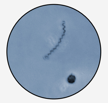
Slide 1
B
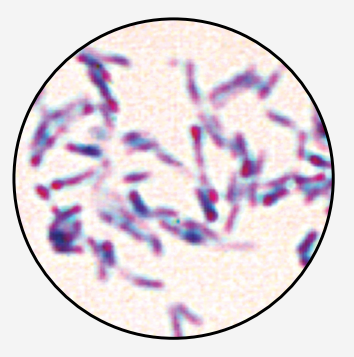
Slide 2
C
Slide 3
D
Slide 4
0 Comments
Verified step by step guidance
1
Step 1: Identify the morphology and staining characteristics of Corynebacterium diphtheriae. C. diphtheriae is a Gram-positive, rod-shaped bacterium that often appears in a characteristic 'club-shaped' or 'palisade' arrangement, sometimes described as resembling Chinese letters. It stains purple with Gram stain due to its thick peptidoglycan cell wall.
Step 2: Examine each slide image for bacterial shape and Gram stain color. Look for purple-stained rods arranged in clusters or palisades, which is typical for C. diphtheriae.
Step 3: Slide 1 shows a spiral-shaped bacterium, which is not consistent with C. diphtheriae morphology.
Step 4: Slide 2 shows purple-stained rod-shaped bacteria arranged in clusters, matching the expected morphology and staining of C. diphtheriae.
Step 5: Slide 3 shows round, red-stained cells, indicating Gram-negative cocci, which are not C. diphtheriae. Slide 4 shows purple-stained cocci, also inconsistent with the rod shape of C. diphtheriae.


 Verified step by step guidance
