Textbook Question
Fill in the following blanks.
a. 1 μm = ______ m
b. 1= _______ 10⁻⁹ m
c. 1 μm = ______ nm
1562
views
 Verified step by step guidance
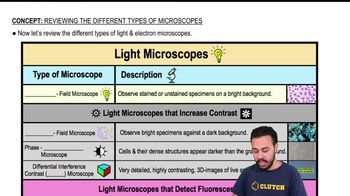
 05:32
05:32 03:52
03:52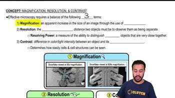 07:29
07:29Fill in the following blanks.
a. 1 μm = ______ m
b. 1= _______ 10⁻⁹ m
c. 1 μm = ______ nm
Assume you stain Bacillus by applying malachite green with heat and then counterstain with safranin. Through the microscope, the green structures are
a. Cell walls
b. Capsules
c. Endospores
d. Flagella
e. Impossible to identify
Three-dimensional images of live cells can be produced with:
a. Darkfield microscopy
b. Fluorescence microscopy
c. Transmission electron microscopy
d. Confocal microscopy
e. Phase-contrast microscopy
Looking at the cell of a photosynthetic microorganism, you observe the chloroplasts are green in brightfield microscopy and red in fluorescence microscopy. You conclude:
a. Chlorophyll is fluorescent
b. The magnification has distorted the image
c. You’re not looking at the same structure in both microscopes
d. The stain masked the green color
e. None of the above
Calculate the total magnification of the nucleus of a cell being observed through a compound light microscope with a 10x ocular lens and an oil immersion lens.
Carbolfuchsin can be used as a simple stain and a negative stain. As a simple stain, the pH is:
a. 2
b. Higher than the negative stain
c. Lower than the negative stain
d. The same as the negative stain