Textbook Question
Explain how the principle, “electrons travel as waves,” applies to microscopy.
1572
views

 Verified step by step guidance
 05:32
05:32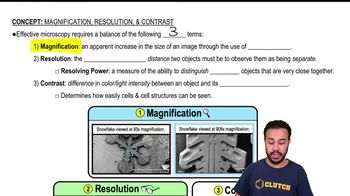 07:29
07:29 02:05
02:05Explain how the principle, “electrons travel as waves,” applies to microscopy.
A nanometer is _______ than a micrometer.
a. 10 times larger
b. 10 times smaller
c. 1000 times larger
d. 1000 times smaller
Label the microscope.
Which of the following is smallest?
a. decimeter
d. millimeter
c. nanometer
d. micrometer
Critique the following definition of magnification given by a student on a microbiology test: “Magnification makes things bigger.”
If an objective magnifies 40× and each binocular lens magnifies 15×, the total magnification of the object being viewed is ________.