Join thousands of students who trust us to help them ace their exams!
Multiple Choice
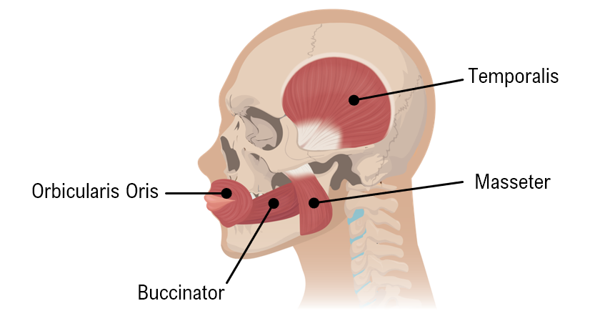
The diagram below shows some of the muscles used for chewing. Using your knowledge of the bones, identify the insertion of the masseter.
A
Temporal Bone.
B
Zygomatic.
C
Mandible.
D
Maxilla.
1 Comment
Verified step by step guidance
1
Identify the masseter muscle in the diagram. It is located on the side of the face, connecting the cheekbone to the lower jaw.
Understand the function of the masseter muscle. It is one of the primary muscles involved in the process of chewing, responsible for elevating the mandible (lower jaw) to close the mouth.
Recall the anatomical terms: 'origin' refers to the fixed attachment, while 'insertion' refers to the movable attachment. For the masseter, the origin is the zygomatic arch (cheekbone).
Determine the insertion point of the masseter muscle. The insertion is where the muscle attaches to the bone that moves when the muscle contracts.
Recognize that the mandible is the bone that moves when the masseter contracts, making it the insertion point of the masseter muscle.


 Verified step by step guidance
