 Back
BackAbdominal Anatomy & Physiology: Ultrasound Study Guide
Study Guide - Smart Notes
 Tailored notes based on your materials, expanded with key definitions, examples, and context.
Introduction to Abdominal Anatomy and Physiology
Overview of Abdominal Imaging
Understanding abdominal anatomy and physiology is essential for interpreting ultrasound images and diagnosing disease. Key aspects include organ location, parenchymal divisions, anatomical landmarks, vasculature, size, appearance, variants, and basic function.
Location: Organs are described in relation to each other and to anatomical planes (anterior/posterior, superior/inferior, medial/lateral).
Parenchymal Divisions: Many organs are divided into functional segments or lobes.
Landmarks: Fissures, ligaments, and vessels help identify and separate organ segments.
Vasculature: Blood supply and drainage are critical for organ function and pathology.
Variants: Anatomical variations can mimic disease or affect function.
Abdominal Cavities and Spaces
Peritoneal and Retroperitoneal Spaces
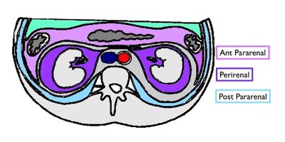
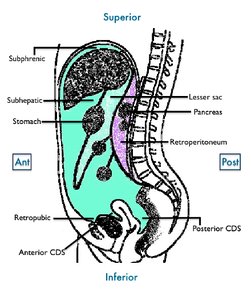
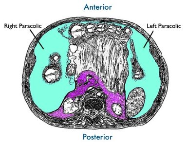
The peritoneum is a closed sac containing major internal organs. Organs are classified as intraperitoneal or retroperitoneal based on their location relative to the peritoneum.
Intraperitoneal: Organs inside the peritoneum (e.g., liver, gallbladder, spleen, stomach, ovaries, some bowel).
Retroperitoneal: Organs beneath the peritoneum, covered anteriorly by peritoneum (e.g., kidneys, adrenal glands, pancreas, duodenum).
Spaces: Subphrenic, subhepatic, lesser sac, paracolic gutters, retropubic, anterior/posterior cul-de-sac (CDS).
Liver Anatomy and Physiology
Liver Structure and Segmentation
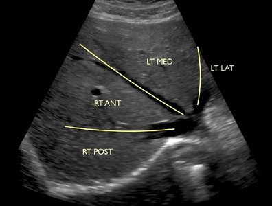
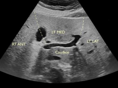
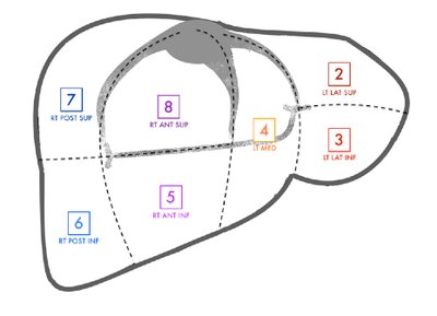
The liver is an intraperitoneal organ (except for the bare area) covered by the Glisson capsule. It is divided into three main lobes: right, left, and caudate. The Couinaud classification system divides the liver into eight segments based on vascular supply and drainage.
Portal Triads: Each segment receives a branch of the portal vein, hepatic artery, and bile duct.
Intersegmental Structures: Hepatic veins, fissures, and ligaments separate segments.
Intrasegmental Structures: Portal veins, bile ducts, and hepatic arteries run within segments.
Liver Vasculature
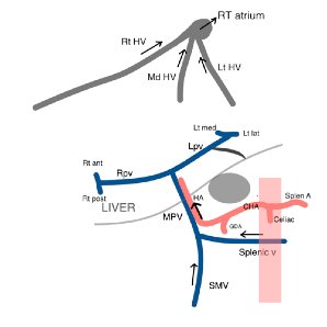
The liver receives blood from the portal vein (70%) and hepatic artery (30%). Blood drains via the hepatic veins into the right atrium.
Portal Vein: Hepatopetal (towards liver), steady flow, minimally phasic.
Hepatic Artery: Hepatopetal, low resistance.
Hepatic Veins: Hepatofugal (away from liver), pulsatile, intersegmental.
Gallbladder and Biliary System
Anatomy and Flow of Bile
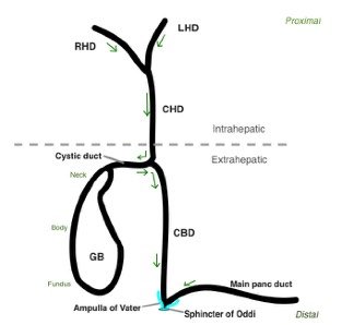
The gallbladder is an intraperitoneal organ that stores and concentrates bile. Bile flows from intrahepatic biliary radicles to the right and left hepatic ducts, then to the common hepatic duct (CHD), cystic duct, gallbladder, common bile duct (CBD), and finally into the duodenum via the ampulla of Vater and sphincter of Oddi.
Wall Layers: Mucosa, fibromuscular, serosa.
Variants: Phrygian cap, Hartmann pouch, junctional fold.
Pancreas Anatomy and Physiology
Pancreatic Structure and Relationships
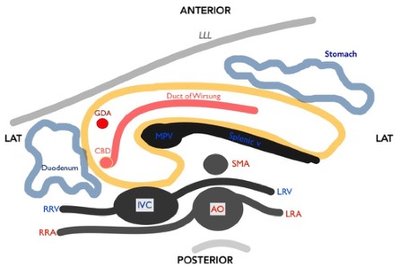
The pancreas is a retroperitoneal organ with both exocrine (enzyme production) and endocrine (hormone production) functions. The main pancreatic duct (duct of Wirsung) joins the CBD at the ampulla of Vater. The accessory duct (duct of Santorini) empties separately into the duodenum.
Vascular Supply: Gastroduodenal artery (head), splenic artery and SMA (body and tail).
Anatomical Relationships: The pancreas is closely related to the IVC, aorta, SMA, splenic vein, and duodenum.
Renal Anatomy and Physiology
Kidney Structure and Blood Flow
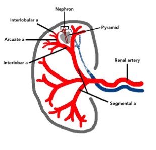
The kidneys are retroperitoneal organs responsible for homeostasis, detoxification, filtration, and urine production. The nephron is the functional unit, located in the cortex. Blood flows through the renal artery, segmental, interlobar, arcuate, and interlobular arteries to the nephron.
Parenchyma: Cortex (outer rim), medulla (pyramids), columns of Bertin.
Sinus: Collecting system (calices, pelvis, ureter).
Summary Table: Key Abdominal Organs and Their Features
Organ | Location | Main Function | Key Vasculature |
|---|---|---|---|
Liver | Intraperitoneal | Metabolism, bile production | Portal vein, hepatic artery, hepatic veins |
Gallbladder | Intraperitoneal | Bile storage | Cystic artery, cystic duct |
Pancreas | Retroperitoneal | Enzyme and hormone production | Gastroduodenal, splenic, SMA |
Kidney | Retroperitoneal | Filtration, urine production | Renal artery, renal vein |
Additional info:
These notes expand on brief points from the original material, providing academic context and definitions for key terms.
Images included are directly relevant to anatomical explanations and reinforce the educational content.
