 Back
BackAdrenal Medulla: Structure, Development, and Function
Study Guide - Smart Notes
 Tailored notes based on your materials, expanded with key definitions, examples, and context.
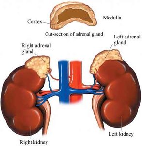
Adrenal Gland: Structure and Location
Overview of the Adrenal Gland
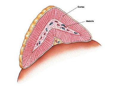
The adrenal glands are paired endocrine organs located on top of the kidneys in mammals. Each gland is composed of two distinct regions: the outer adrenal cortex and the inner adrenal medulla. These regions are functionally and developmentally separate, with unique embryological origins and physiological roles.
Adrenal Cortex: The outer layer, responsible for steroid hormone production.
Adrenal Medulla: The central region, specialized for catecholamine synthesis and release.
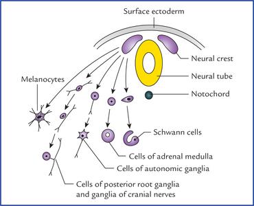
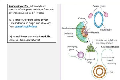
Developmental Origins of the Adrenal Medulla
Embryology and Morphology
The adrenal cortex and medulla have distinct embryological origins. The cortex arises from mesodermal tissue, while the medulla is derived from the neural crest (ectoderm). The adrenal medulla is functionally and developmentally part of the sympathetic nervous system, acting as a modified sympathetic ganglion.
Neural Crest Derivation: Pheochromoblasts migrate from the neural crest and differentiate into chromaffin cells (modified neuronal cells).
Sympathetic Integration: The adrenal medulla reinforces the actions of the sympathetic nervous system by releasing catecholamines during stress.
Cell Types of the Adrenal Medulla
Chromaffin Cells and Sympathetic Ganglion Cells
The adrenal medulla contains two main cell types:
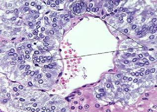
Pheochromocytes (Chromaffin Cells): Axonless secretory cells containing granules of catecholamines, ATP, proteins, and lipids. These cells stain brown with chromate due to their catecholamine content.
Sympathetic Ganglion Cells: True neuronal cells, present in small numbers.
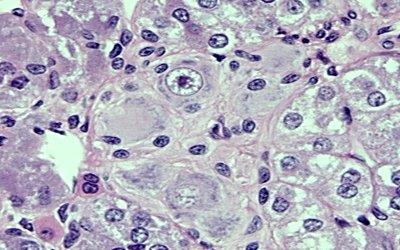
Chromaffin cells are further classified based on the catecholamine they store:
Epinephrine (Adrenaline) Storing Cells: Numerous, weak chromate staining, small and less dense granules.
Norepinephrine (Noradrenaline) Storing Cells: Fewer, strong chromate staining, large dense granules.
Distribution of Chromaffin Cells
Locations Beyond the Adrenal Medulla
Chromaffin cells are not exclusive to the adrenal medulla. They are also found in other tissues such as the carotid bodies, liver, heart, kidney, and gonads in mammals and birds. These cells are responsible for catecholamine production in these locations.
Species Variation: In some vertebrates, the association between chromaffin tissue and adrenal steroidogenic tissue varies, influencing the production of norepinephrine (NE) versus epinephrine (E).
Regulation of Catecholamine Synthesis
Glucocorticoid Influence and PNMT
The synthesis of epinephrine in chromaffin cells is regulated by glucocorticoids produced in the adrenal cortex. During early development, chromaffin cells produce only norepinephrine. The enzyme phenylethanolamine N-methyltransferase (PNMT), which converts NE to E, is induced by glucocorticoids.
PNMT: Key enzyme for epinephrine synthesis, regulated by glucocorticoids.
Developmental Regulation: In mammals, the proximity of the cortex and medulla ensures high glucocorticoid exposure, promoting epinephrine production.
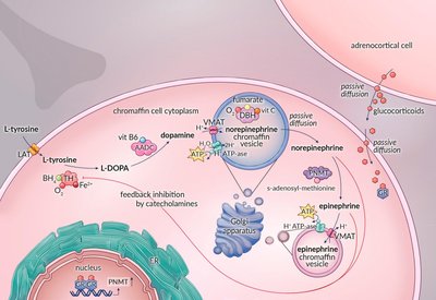
Catecholamine Biosynthesis Pathway
Steps in Catecholamine Synthesis
Catecholamines are synthesized from the amino acid tyrosine through a series of enzymatic reactions:
Tyrosine is converted to L-DOPA.
L-DOPA is converted to dopamine.
Dopamine is converted to norepinephrine (NE).
NE is converted to epinephrine (E) by PNMT.
Catecholamines are stored in secretory granules and released in response to sympathetic stimulation, allowing rapid response to environmental stressors.
Proportion Released: Approximately 80% of released catecholamines are epinephrine.
Mechanism of Action: Catecholamine Receptors
Adrenergic Receptor Subtypes and Effects
Catecholamines exert their effects by binding to adrenergic receptors, which are G protein-coupled receptors. There are at least nine subtypes, classified as alpha (α) and beta (β) receptors. The physiological response depends on the receptor subtype and its distribution in target tissues.
Cardiac Stimulation: Epinephrine (E) is more potent than norepinephrine (NE) at stimulating cardiac output (β-receptor mediated).
Vasoconstriction: NE is more potent than E at causing blood vessel constriction (α-receptor mediated).
Metabolic Rate: E increases metabolic rate 5–10 times more than NE.
Receptor | Norepinephrine | Epinephrine |
|---|---|---|
α (alpha) | +++++ | ++++ |
β (beta) | ++ | ++++ |
Pheochromocytoma: Pathophysiology and Clinical Features
Chromaffin Cell Tumors
Pheochromocytoma is a tumor of chromaffin cells, most commonly arising in the adrenal medulla (90%), but can also occur extra-adrenally (paraganglioma). These tumors secrete excessive catecholamines, leading to characteristic clinical symptoms.
Signs and Symptoms: Treatment-resistant hypertension (95%), headache, sweating, palpitations, chest pain, anxiety, glucose intolerance, and increased metabolic rate. The classic triad includes headache, sweating, and palpitations.
Diagnosis: Elevated plasma catecholamines and increased urinary metabolites.
Treatment: Surgical resection is the mainstay. Preoperative management includes non-specific and irreversible alpha-adrenoceptor blockade (e.g., phenoxybenzamine) to prevent hypertensive crises.
Summary Table: Key Features of the Adrenal Medulla
Feature | Description |
|---|---|
Location | Central region of adrenal gland, above kidneys |
Cell Types | Chromaffin cells (epinephrine and norepinephrine storing), sympathetic ganglion cells |
Developmental Origin | Neural crest (ectoderm) |
Main Hormones | Epinephrine (80%), Norepinephrine (20%) |
Regulation | Sympathetic stimulation, glucocorticoids (for E synthesis) |
Clinical Condition | Pheochromocytoma (chromaffin cell tumor) |
