 Back
BackAnatomy of Blood Vessels and Circulatory Pathways
Study Guide - Smart Notes
 Tailored notes based on your materials, expanded with key definitions, examples, and context.
Cardiovascular System Overview
Components of the Cardiovascular System
The cardiovascular system is responsible for the transport of blood, nutrients, gases, and wastes throughout the body. It consists of the heart and an extensive network of blood vessels that form two main circuits:
Pulmonary Circuit: Carries blood between the heart and lungs for gas exchange.
Systemic Circuit: Delivers oxygenated blood from the heart to the rest of the body and returns deoxygenated blood back to the heart.
Histological Organization of Blood Vessels
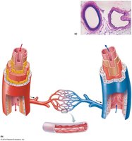
Structure of Vessel Walls
Blood vessels are composed of three distinct layers, each with specialized functions:
Tunica intima: The innermost layer, consisting of endothelium and a subendothelial layer, providing a smooth surface for blood flow.
Tunica media: The middle layer, primarily made of smooth muscle and elastic fibers, responsible for vasoconstriction and vasodilation.
Tunica externa (adventitia): The outermost layer, composed of connective tissue, providing structural support and anchoring the vessel to surrounding tissues.
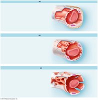
Arteries vs. Veins
Key Differences
Arteries:
Carry blood away from the heart (usually oxygenated, except pulmonary arteries).
Thicker tunica media for higher pressure resistance.
Smaller lumen compared to veins.
No valves (except in the pulmonary trunk and aorta).
Elastic and muscular types based on function and location.
Veins:
Carry blood toward the heart (usually deoxygenated, except pulmonary veins).
Thinner tunica media and larger lumen.
Contain valves, especially in limbs, to prevent backflow.
Serve as blood reservoirs.
Types of Arteries
Elastic (Conducting) Arteries
Largest arteries (e.g., aorta, pulmonary trunk).
High content of elastic fibers allows them to stretch and recoil with each heartbeat.
Help maintain continuous blood flow during diastole.
Muscular (Distributing) Arteries
Medium-sized arteries (e.g., radial, femoral arteries).
Thicker tunica media with more smooth muscle.
Regulate blood flow to specific organs via vasoconstriction and vasodilation, controlled by the sympathetic nervous system (SNS).
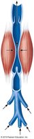
Arterioles
Arterioles are the smallest branches of arteries leading to capillaries. They play a crucial role in regulating blood pressure and flow into capillary beds by adjusting their diameter.
Major site of vascular resistance.
Controlled by local factors and autonomic nervous system.
Capillaries
Structure and Function
Capillaries are the smallest and most delicate blood vessels, serving as the primary site for exchange of gases, nutrients, and wastes between blood and tissues.
Single layer of endothelial cells for efficient exchange.
Three main types:
Continuous capillaries: Most common; uninterrupted endothelium (e.g., muscle, skin).
Fenestrated capillaries: Have pores for increased permeability (e.g., kidneys, intestines).
Sinusoids: Large gaps for passage of cells and large molecules (e.g., liver, spleen).
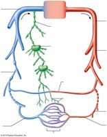
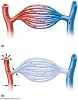
Capillary Beds (Capillary Plexus)
Capillaries form interconnected networks called capillary beds or plexuses, which regulate blood flow through tissues.
Precapillary sphincters control entry of blood into capillaries.
Thoroughfare channels allow direct flow from arterioles to venules.
Veins
Types of Veins
Venules: Smallest veins, collect blood from capillaries.
Medium-sized veins: Contain valves, especially in limbs, to prevent backflow.
Large veins: Include the superior and inferior vena cava, returning blood directly to the heart.
Venous Valves
Valves in veins, especially in the extremities, prevent the backflow of blood and aid in venous return to the heart, particularly against gravity.
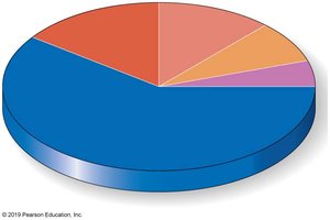
Blood Distribution in the Body
Blood is unevenly distributed in the body, with the majority found in the venous system, which acts as a reservoir.
About 60-70% of blood volume is in the veins at rest.
Arteries and capillaries contain the remainder.
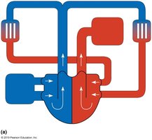
Blood Circulation Pathways
Pulmonary Circuit
The pulmonary circuit carries deoxygenated blood from the right ventricle of the heart to the lungs and returns oxygenated blood to the left atrium.
Right ventricle → Pulmonary arteries → Lungs → Pulmonary veins → Left atrium
Systemic Circuit
The systemic circuit delivers oxygenated blood from the left ventricle to the body and returns deoxygenated blood to the right atrium.
Left ventricle → Aorta → Body tissues → Veins → Right atrium
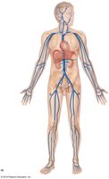
Major Arteries of the Body
The major arteries branch from the aorta and supply oxygenated blood to all regions of the body.
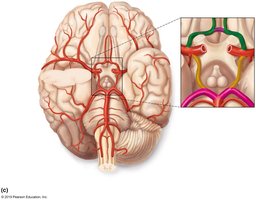
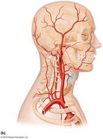
Major Arteries of the Head and Neck
Key arteries include the common carotid, internal and external carotid, and vertebral arteries, which supply the brain and facial structures.
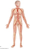
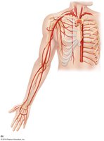
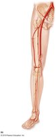
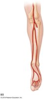
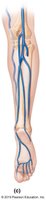
Major Arteries of the Extremities
Arteries of the arms and legs include the subclavian, axillary, brachial, radial, ulnar, femoral, popliteal, anterior tibial, and posterior tibial arteries.
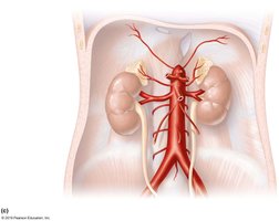
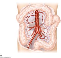
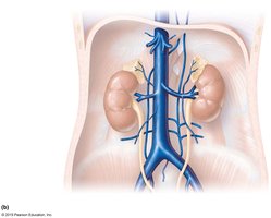
Major Arteries of the Thorax and Abdomen
These include the thoracic aorta, abdominal aorta, celiac trunk, superior and inferior mesenteric arteries, and renal arteries.

Major Veins of the Body
Major veins return deoxygenated blood to the heart. The superior and inferior vena cava are the largest veins, draining the upper and lower body, respectively.
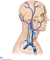
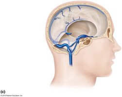
Major Veins of the Head and Neck
Important veins include the internal and external jugular veins, and the dural venous sinuses of the brain.
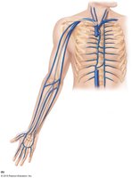
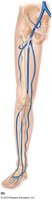
Major Veins of the Extremities
Veins of the arms and legs include the subclavian, axillary, cephalic, basilic, brachial, femoral, great saphenous, and small saphenous veins.
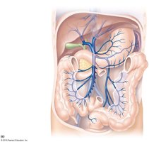
Major Veins of the Thorax and Abdomen
These include the hepatic portal vein, renal veins, and the inferior vena cava, which collects blood from the lower body and abdominal organs.
Summary Table: Comparison of Arteries, Veins, and Capillaries
Feature | Arteries | Veins | Capillaries |
|---|---|---|---|
Wall Thickness | Thick | Thin | Very thin (one cell layer) |
Lumen Size | Narrow | Wide | Very narrow |
Valves | Absent | Present (especially in limbs) | Absent |
Function | Carry blood away from heart | Carry blood toward heart | Exchange of substances |
Pressure | High | Low | Very low |
