 Back
BackAnatomy of the Eye: Structure, Function, and Visual Pathways
Study Guide - Smart Notes
 Tailored notes based on your materials, expanded with key definitions, examples, and context.
Anatomy of the Eye
Introduction
The eye is a highly specialized sensory organ responsible for vision. It is composed of several structural components and accessory structures that work together to capture and process visual information. Understanding the anatomy of the eye is essential for comprehending how vision occurs and how the eye adapts to different visual environments.
Accessory Structures of the Eye
Lacrimal Apparatus
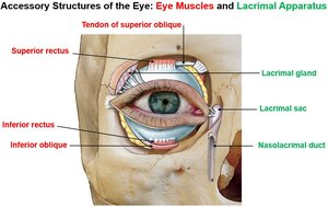
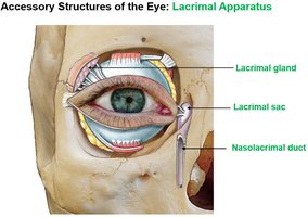
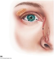
The lacrimal apparatus is responsible for the production, distribution, and drainage of tears, which lubricate and protect the eye.
Lacrimal gland: Produces tears containing lysozyme, which lubricates the eye and provides a germicidal effect.
Lacrimal sac and nasolacrimal duct: Drain tears from the eye surface into the nasal cavity.
Extrinsic Eye Muscles
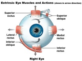
Six extrinsic (skeletal) muscles control the movement of the eyeball, allowing for precise positioning and tracking of objects. These muscles are innervated by cranial nerves III (oculomotor), IV (trochlear), and VI (abducens).
Superior rectus – Elevates the eye
Inferior rectus – Depresses the eye
Lateral rectus – Abducts the eye (moves it laterally)
Medial rectus – Adducts the eye (moves it medially)
Superior oblique – Depresses and medially rotates the eye
Inferior oblique – Elevates and laterally rotates the eye
Muscle | Nerve Innervation | Action when contracting |
|---|---|---|
Lateral rectus | CN VI - Abducens nerve | Abduction |
Medial rectus | CN III - Oculomotor nerve | Adduction |
Superior rectus | CN III - Oculomotor nerve | Elevation |
Inferior rectus | CN III - Oculomotor nerve | Depression |
Inferior oblique | CN III - Oculomotor nerve | Elevation and lateral rotation |
Superior oblique | CN IV - Trochlear nerve | Depression and medial rotation |
Intrinsic Eye Muscles
Intrinsic muscles are smooth muscles located within the eye, responsible for controlling lens shape and pupil size.
Ciliary muscle: Adjusts the curvature of the lens for focusing on near or distant objects (accommodation).
Pupillary muscles (sphincter and dilator): Control the size of the pupil to regulate the amount of light entering the eye.
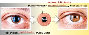
Pupillary light reflex: The pupil constricts in response to bright light (parasympathetic stimulation) and dilates in dim light (sympathetic stimulation).
Structural Components of the Eye
Wall Layers (Tunics) of the Eyeball
The wall of the eyeball consists of three main layers, each with distinct functions:
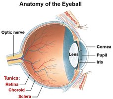
Sclera: The outermost fibrous layer, known as the "white" of the eye, provides protection and shape.
Choroid: The middle vascular layer, rich in blood vessels, supplies nutrients and oxygen to the retina.
Retina: The innermost neural layer, containing photoreceptors responsible for detecting light.
Major Parts of the Eye
Cornea: Transparent anterior portion that refracts light entering the eye.
Iris: Colored part of the eye containing pupillary muscles to control pupil size.
Pupil: Central opening in the iris through which light passes.
Lens: Biconvex structure that focuses light onto the retina; its shape is adjusted by the ciliary muscle via the suspensory ligament.
Ciliary body: Contains the ciliary muscle and produces aqueous humor.
Suspensory ligament: Connects the ciliary body to the lens, enabling lens shape changes.
Internal Cavities of the Eye
The interior of the eye is divided into two main cavities by the lens and ciliary body:
Anterior cavity: Located in front of the lens, further divided by the iris into the anterior chamber (between cornea and iris) and posterior chamber (between iris and lens). Filled with aqueous humor, which maintains intraocular pressure and nourishes avascular structures.
Posterior cavity: Located behind the lens, filled with vitreous humor (gel-like substance) that helps maintain the shape of the eye and holds the retina in place.
Retina: Structure and Function
Cellular Layers of the Retina
The retina is composed of three main cellular layers, each with specialized functions in visual processing:
Ganglion cell layer: Contains ganglion cells whose axons form the optic nerve (CN II).
Bipolar cell layer: Intermediate layer that transmits signals from photoreceptors to ganglion cells.
Photoreceptor layer: Contains rods and cones, the sensory receptors for vision.
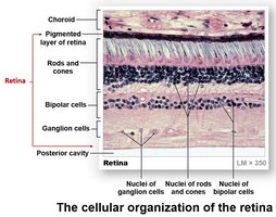
Rods: Specialized for scotopic (low-light) vision; more numerous in the peripheral retina.
Cones: Responsible for color vision and high visual acuity; concentrated in the macula lutea, especially the fovea centralis.
Specific Regions of the Retina
Fovea centralis: A small pit in the center of the macula lutea with the highest density of cones; site of sharpest vision and color discrimination.
Optic disc: The point where ganglion cell axons exit the eye to form the optic nerve; lacks photoreceptors and is known as the "blind spot."
Visual Pathway
Major Components
The visual pathway transmits visual information from the retina to the brain for processing and interpretation.
Optic nerve (CN II): Formed by axons of ganglion cells, carries visual signals from the retina.
Optic chiasm: Site where some optic nerve fibers cross to the opposite side of the brain.
Optic tract: Carries visual information from the optic chiasm to the lateral geniculate nucleus of the thalamus.
Visual cortex: Located in the occipital lobe, it is the primary center for processing visual information.
Summary
All structures of the eye work together to enable vision. The eye adjusts the amount of light entering by changing pupil size, focuses images using the cornea and lens, converts light into electrical signals via photoreceptors, and transmits these signals through the visual pathway to the brain, where they are interpreted as images.
