 Back
BackArticulations (Joints): Structure, Classification, and Movements
Study Guide - Smart Notes
 Tailored notes based on your materials, expanded with key definitions, examples, and context.
Articulations (Joints)
Introduction to Joints
Joints, or articulations, are the sites where two or more bones meet. They are the weakest parts of the skeleton but are essential for providing mobility and holding the skeleton together.
Articulation: The location at which bones connect.
Functions of joints:
Provide mobility to the skeleton
Hold the skeleton together
Classification of Joints
Structural Classification
Structural classification is based on the material binding the bones and the presence or absence of a joint cavity. The three main types are:
Fibrous joints – Bones joined by fibrous tissue; no joint cavity; mostly immovable.
Cartilaginous joints – Bones united by cartilage; no joint cavity; variable mobility.
Synovial joints – Bones separated by a fluid-filled joint cavity; freely movable.
Functional Classification
Functional classification is based on the degree of movement allowed:
Synarthroses – Immovable joints
Amphiarthroses – Slightly movable joints
Diarthroses – Freely movable joints
Fibrous Joints
General Features
Fibrous joints are connected by dense connective tissue and lack a joint cavity. Most are immovable.
Sutures: Found between skull bones; interlocking junctions filled with connective tissue fibers. Allow growth during youth; fuse in adulthood (synostoses).
Syndesmoses: Bones connected by ligaments; movement varies from immovable to slightly movable (e.g., tibia-fibula, radius-ulna).
Gomphoses: Peg-in-socket joints (e.g., tooth in alveolar socket); connected by the periodontal ligament.
Cartilaginous Joints
General Features
Cartilaginous joints unite bones with cartilage and lack a joint cavity. Two main types:
Synchondroses: Bones united by hyaline cartilage; all are immovable (synarthrotic). Examples: epiphyseal plates, first rib-sternum joint.
Symphyses: Articulating surfaces covered with hyaline cartilage fused to a pad of fibrocartilage; amphiarthrotic (slightly movable). Examples: intervertebral joints, pubic symphysis.
Synovial Joints
General Structure
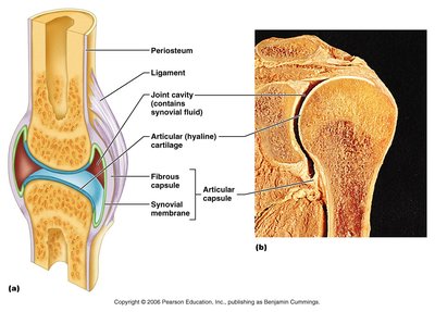
Synovial joints are characterized by a fluid-filled joint cavity and are all freely movable (diarthroses). They are the most common type of joint in the body.
Articular cartilage: Hyaline cartilage covering bone ends, reducing friction.
Joint (synovial) cavity: Space containing synovial fluid.
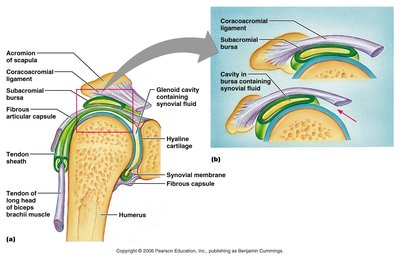
Articular capsule: Double-layered capsule enclosing the joint cavity (outer fibrous layer and inner synovial membrane).
Synovial fluid: Lubricates and nourishes articular cartilage.
Reinforcing ligaments: Strengthen and support the joint.
Friction-Reducing Structures
Bursae: Flattened sacs lined with synovial membrane, containing synovial fluid; reduce friction where structures rub together.
Tendon sheath: Elongated bursa that wraps around a tendon, especially in the digits.
Stability of Synovial Joints
Articular surfaces: Shape determines possible movements.
Ligaments: Unite bones and prevent excessive movement.
Muscle tendons: Cross joints and help stabilize them.
Range of Motion
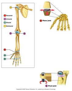
Nonaxial: Slipping movements only
Uniaxial: Movement in one plane
Biaxial: Movement in two planes
Multiaxial: Movement in or around all three planes
Types of Synovial Joints
Plane Joints
Articular surfaces are essentially flat, allowing only slipping or gliding movements. These are the only examples of nonaxial joints. Example: intercarpal joints.
Hinge Joints
Cylindrical projection of one bone fits into a trough-shaped surface of another. Motion is along a single plane (uniaxial), permitting flexion and extension only. Examples: elbow, interphalangeal joints.
Pivot Joints
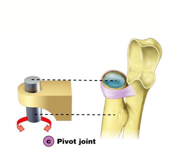
Rounded end of one bone protrudes into a ring formed by another bone and ligament. Only uniaxial movement is allowed. Examples: joint between the atlas and dens (atlantoaxial joint), proximal radioulnar joint.
Condyloid (Ellipsoidal) Joints
Oval articular surface of one bone fits into a complementary depression in another. Biaxial joints permit all angular motions. Examples: radiocarpal (wrist) joints, metacarpophalangeal (knuckle) joints.
Saddle Joints
Similar to condyloid joints but allow greater movement. Each articular surface has both concave and convex areas. Example: carpometacarpal joint of the thumb.
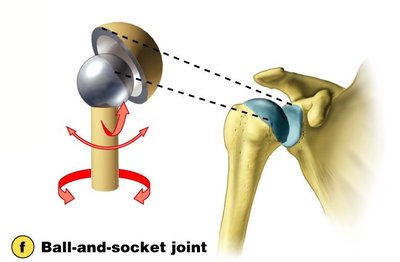
Ball-and-Socket Joints
Spherical head of one bone articulates with a cuplike socket of another. Multiaxial joints permit the most freely moving synovial joints. Examples: shoulder and hip joints.
Movements at Synovial Joints
Gliding Movements
One flat bone surface glides or slips over another. Examples: intercarpal and intertarsal joints, articular processes of vertebrae.
Angular Movements
Flexion: Decreases the angle of the joint.
Extension: Increases the angle of the joint.
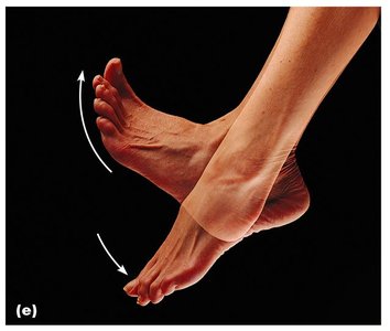
Dorsiflexion and plantar flexion: Up and down movement of the foot at the ankle.
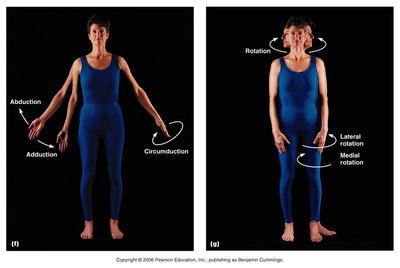
Abduction: Movement away from the midline.
Adduction: Movement toward the midline.
Circumduction: Movement describes a cone in space (combination of flexion, abduction, extension, and adduction).
Rotation
Turning of a bone around its own long axis. Examples: between first two vertebrae, hip and shoulder joints.
Special Movements
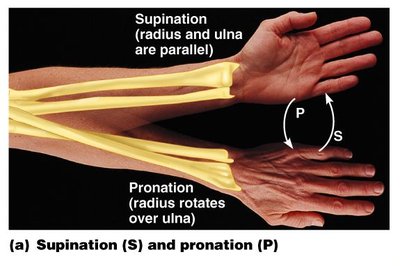
Supination and pronation: Rotational movement of the forearm; supination turns the palm up, pronation turns it down.
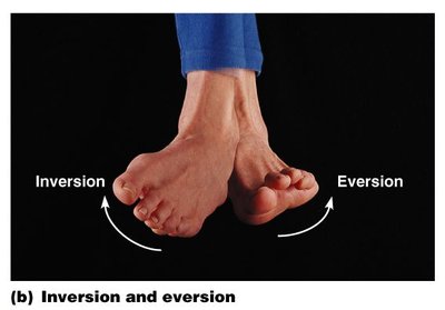
Inversion and eversion: Movements of the sole of the foot inward (inversion) or outward (eversion).
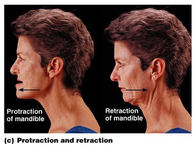
Protraction and retraction: Anterior and posterior movement in a transverse plane (e.g., mandible).
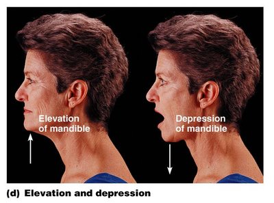
Elevation and depression: Lifting a body part superiorly or moving it inferiorly (e.g., mandible).
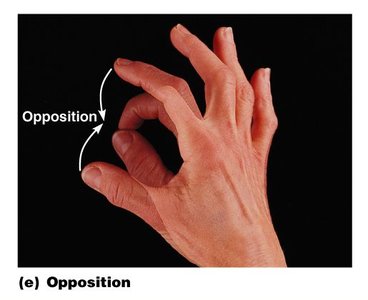
Opposition: Movement of the thumb to touch the tips of other fingers.
Summary Table: Types of Joints and Movements
Joint Type | Structural Feature | Movement | Example |
|---|---|---|---|
Fibrous (Suture) | Interlocking bones with connective tissue | Immovable | Skull sutures |
Fibrous (Syndesmosis) | Ligament connection | Slightly movable | Distal tibiofibular joint |
Fibrous (Gomphosis) | Peg-in-socket | Immovable | Tooth in socket |
Cartilaginous (Synchondrosis) | Hyaline cartilage | Immovable | Epiphyseal plate |
Cartilaginous (Symphysis) | Fibrocartilage pad | Slightly movable | Pubic symphysis |
Synovial (Plane) | Flat surfaces | Gliding | Intercarpal joints |
Synovial (Hinge) | Cylinder and trough | Flexion/Extension | Elbow |
Synovial (Pivot) | Rounded bone in ring | Rotation | Proximal radioulnar joint |
Synovial (Condyloid) | Oval surfaces | All angular | Wrist |
Synovial (Saddle) | Concave/convex | Greater movement | Thumb |
Synovial (Ball-and-Socket) | Spherical head/socket | Multiaxial | Shoulder, hip |
Additional info: The formal name for the joint between the atlas and the dens is the atlantoaxial joint. The joint between the occiput and the atlas is the atlanto-occipital joint.
