 Back
BackArticulations (Joints): Structure, Function, and Clinical Relevance
Study Guide - Smart Notes
 Tailored notes based on your materials, expanded with key definitions, examples, and context.
Chapter 8: Articulations (Joints)
Introduction to Joints (Articulations)
Joints, or articulations, are the locations where two or more bones meet. They are essential for movement, stability, and skeletal growth. The structure and function of joints determine the range and type of motion possible in the human body.
Functions of Joints
Enable Movement: Muscles and tendons exert force across joints, allowing for a wide variety of movements.
Provide Stability: Joints with limited or no movement (e.g., skull sutures) are highly stable and protect underlying structures.
Allow Bone Growth: The epiphyseal plate (a temporary joint) enables long bones to lengthen during development.
Classification of Joints
Functional Classification
Synarthrosis: Immovable joints; provide maximum stability (e.g., skull sutures).
Amphiarthrosis: Slightly movable joints; offer both stability and limited movement (e.g., intervertebral discs).
Diarthrosis: Freely movable joints; allow a wide range of motion but less stability (e.g., shoulder joint).
Structural Classification
Fibrous Joints: Bones united by dense regular collagenous connective tissue; no joint cavity; mostly synarthroses or amphiarthroses.
Cartilaginous Joints: Bones joined by cartilage; no joint cavity; functionally synarthroses or amphiarthroses.
Synovial Joints: Bones separated by a fluid-filled joint cavity; always diarthroses.
Fibrous Joints
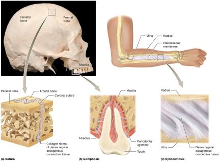
Fibrous joints are connected by dense collagen fibers, providing stability and permitting little to no movement.
Sutures: Found between skull bones; interlocking projections provide strong, immovable joints. Fusion later in life forms a synostosis.
Gomphoses: Joints between teeth and their sockets (alveoli) in the mandible or maxilla, stabilized by the periodontal ligament.
Syndesmoses: Bones joined by an interosseous membrane or ligament (e.g., between radius and ulna); allow limited movement.
Cartilaginous Joints
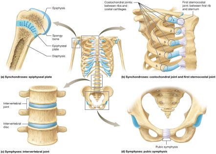
Cartilaginous joints lack a joint cavity and allow little movement. They are classified based on the type of cartilage present.
Synchondroses: Bones united by hyaline cartilage; immovable (e.g., epiphyseal plate, first sternocostal joint).
Symphyses: Bones united by a fibrocartilage pad; slightly movable (e.g., intervertebral discs, pubic symphysis).
Clinical Note: Epiphyseal Plate Fractures
Epiphyseal plates are weak points in growing bones and susceptible to fractures, which can lead to limb length discrepancies or deformities if not properly managed.
Common in children due to recreational activities or sports.
Synovial Joints
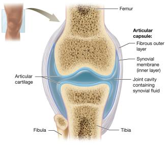
Synovial joints are the most common and movable type of joint in the body. They are characterized by a joint cavity filled with synovial fluid.
Articular Capsule: Double-layered; outer fibrous layer provides strength, inner synovial membrane secretes synovial fluid.
Synovial Fluid: Lubricates, nourishes, and absorbs shock within the joint.
Articular Cartilage: Hyaline cartilage covers bone surfaces, reducing friction and absorbing shock.
Other Components: Adipose tissue for padding, blood vessels for nutrient supply, and nerves for sensation.
Stabilizing and Supportive Structures
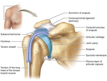
Ligaments: Connect bone to bone; can be intrinsic (within the capsule) or extrinsic (outside the capsule).
Tendons: Connect muscle to bone; stabilize joints by maintaining muscle tone.
Bursae: Fluid-filled sacs that reduce friction in areas of high stress.
Tendon Sheaths: Elongated bursae that protect tendons in high-friction regions.
Clinical Note: Bursitis and Arthritis
Bursitis: Inflammation of a bursa, often due to trauma or repetitive motion; treated with rest, ice, and anti-inflammatory medications.
Arthritis: Inflammation of joints, leading to pain and reduced mobility. Types include osteoarthritis (wear and tear), rheumatoid arthritis (autoimmune), and gouty arthritis (uric acid crystals).
Functional Classes of Synovial Joints
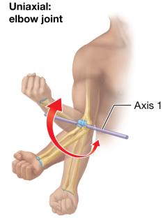
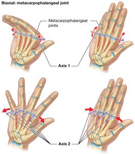
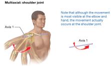
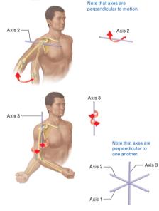
Synovial joints are classified by the number of axes around which bones can move:
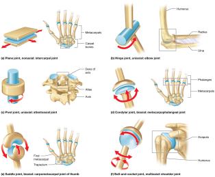
Nonaxial: Movement in one or more planes, but not around an axis (e.g., intercarpal joints).
Uniaxial: Movement around one axis (e.g., elbow joint).
Biaxial: Movement around two axes (e.g., metacarpophalangeal joints).
Multiaxial (Triaxial): Movement around three axes (e.g., shoulder joint).
Movements at Synovial Joints
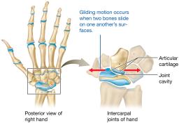
Gliding Movements
Sliding motion between flat surfaces of bones; nonaxial (e.g., intercarpal joints).
Angular Movements
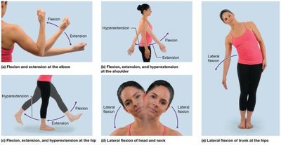
Flexion: Decreases the angle between bones (e.g., bending the elbow).
Extension: Increases the angle between bones.
Hyperextension: Extension beyond anatomical position.
Lateral Flexion: Sideways movement (e.g., bending the trunk).
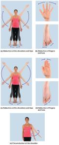
Abduction: Movement away from the midline.
Adduction: Movement toward the midline.
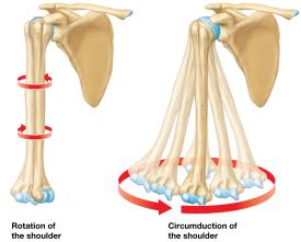
Circumduction: Circular movement combining flexion, extension, abduction, and adduction.
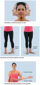
Rotation: Bone pivots around its own longitudinal axis.
Internal (Medial) Rotation: Toward the midline.
External (Lateral) Rotation: Away from the midline.
Special Movements
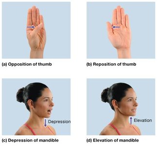
Opposition/Reposition: Movement of the thumb across the palm and back.
Depression/Elevation: Inferior and superior movement (e.g., opening and closing the mouth).
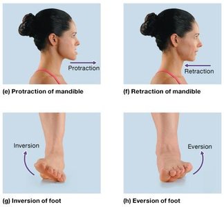
Protraction/Retraction: Anterior and posterior movement (e.g., jutting jaw forward and back).
Inversion/Eversion: Rotational movement of the foot inward or outward.
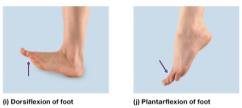
Dorsiflexion/Plantarflexion: Movement of the foot upward (toes toward head) or downward (toes toward ground).
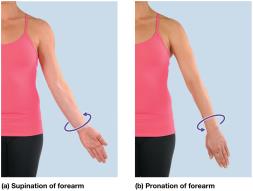
Supination/Pronation: Rotation of the forearm so the palm faces anteriorly (supination) or posteriorly (pronation).
Range of Motion
Nonaxial joints have the smallest range of motion; multiaxial joints have the greatest.
Structural Classes of Synovial Joints
The shape of articulating surfaces and the number of axes determine the type of synovial joint and its range of motion.
Type | Axes | Example |
|---|---|---|
Plane | Nonaxial | Intercarpal joints |
Hinge | Uniaxial | Elbow joint |
Pivot | Uniaxial | Atlantoaxial joint |
Condylar (Ellipsoid) | Biaxial | Metacarpophalangeal joints |
Saddle | Biaxial | Thumb carpometacarpal joint |
Ball-and-Socket | Multiaxial | Shoulder, hip joints |
Specific Hinge Joints: The Elbow and the Knee
The Elbow Joint
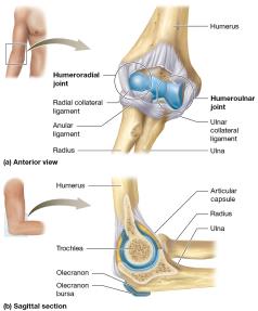
Composed of the humeroulnar and humeroradial joints.
Stabilized by the radial (lateral) collateral, ulnar (medial) collateral, and anular ligaments.
The Knee Joint
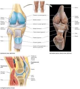
Largest diarthrosis; hinge joint with some rotation and gliding.
Composed of tibiofemoral and patellofemoral joints.
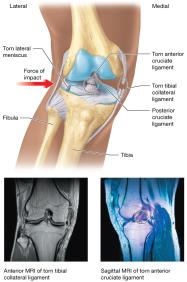
Stabilized by menisci, collateral ligaments, and cruciate ligaments (ACL and PCL).
Knee Injuries: The Unhappy Triad
Lateral blows to the knee can rupture the tibial collateral ligament, lateral meniscus, and anterior cruciate ligament.
Common in contact sports; often requires surgical repair.
Specific Ball-and-Socket Joints: The Shoulder and the Hip
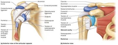
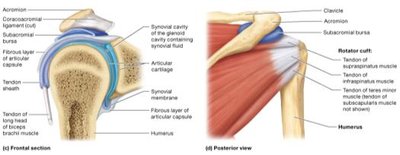
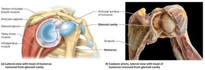
The Shoulder (Glenohumeral Joint)
Most freely movable joint; less stable.
Stabilized by the articular capsule, rotator cuff muscles, glenoid labrum, and several ligaments and bursae.
Shoulder Dislocations
Most dislocations occur anteriorly and inferiorly due to the weakness of the capsule in this region.
Separated shoulder involves the acromioclavicular joint, not the glenohumeral joint.
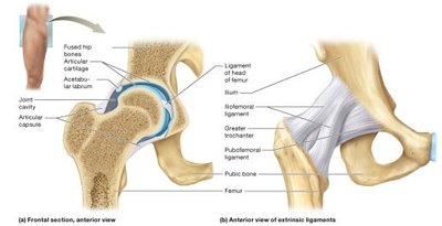
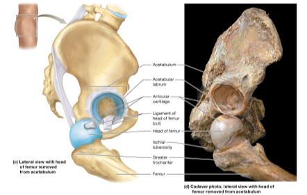
The Hip (Coxal Joint)
Ball-and-socket joint between the acetabulum and femoral head; more stable than the shoulder.
Stabilized by the acetabular labrum, strong articular capsule, and several large ligaments (iliofemoral, ischiofemoral, pubofemoral, ligament of the head of femur).
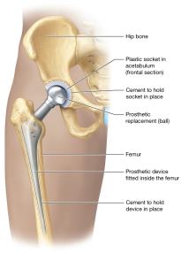
Hip Joint Replacement Surgery
Indicated for severe arthritis, trauma, or bone tumors.
Total replacement involves both the femoral head and acetabulum; partial replacement involves only the femoral head.
Physical therapy is essential for recovery.
Summary Table: Joint Types and Examples
Joint Type | Structural Classification | Functional Classification | Example |
|---|---|---|---|
Suture | Fibrous | Synarthrosis | Skull |
Gomphosis | Fibrous | Synarthrosis | Tooth in socket |
Syndesmosis | Fibrous | Amphiarthrosis | Radius and ulna |
Synchondrosis | Cartilaginous | Synarthrosis | Epiphyseal plate |
Symphysis | Cartilaginous | Amphiarthrosis | Pubic symphysis |
Plane | Synovial | Diarthrosis | Intercarpal joints |
Hinge | Synovial | Diarthrosis | Elbow, knee |
Pivot | Synovial | Diarthrosis | Atlantoaxial joint |
Condylar | Synovial | Diarthrosis | Metacarpophalangeal joints |
Saddle | Synovial | Diarthrosis | Thumb joint |
Ball-and-Socket | Synovial | Diarthrosis | Shoulder, hip |
Additional info: This guide covers the essential structure, function, and clinical relevance of joints (articulations) as outlined in a typical Anatomy & Physiology curriculum. For further study, refer to clinical cases and laboratory exercises related to joint injuries and pathologies.
