 Back
BackAutonomic Nervous System: Structure, Function, and Divisions
Study Guide - Smart Notes
 Tailored notes based on your materials, expanded with key definitions, examples, and context.
The Autonomic Nervous System (ANS)
Introduction to the ANS
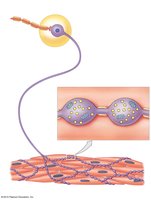
The autonomic nervous system (ANS) is a critical component of the peripheral nervous system that operates largely outside conscious awareness. It regulates essential involuntary functions, including body temperature, cardiovascular, respiratory, digestive, excretory, and reproductive systems.
Regulation: Maintains homeostasis by adjusting internal organ activity.
Coordination: Integrates multiple organ systems for efficient function.
Comparison of Somatic and Autonomic Nervous Systems
Somatic vs. Autonomic Pathways
The somatic and autonomic nervous systems differ in their targets and pathways:
Somatic Nervous System: Innervates skeletal muscles; afferent pathways originate in skeletal muscle receptors.
Autonomic Nervous System: Innervates visceral organs; afferent pathways originate in visceral receptors.
Divisions of the Autonomic Nervous System
Sympathetic and Parasympathetic Subdivisions
The ANS is divided into two major subdivisions:
Sympathetic Division (Thoracolumbar): Most active during stress, exertion, or emergencies; prepares the body for "fight or flight" responses.
Parasympathetic Division (Craniosacral): Most active during resting conditions; promotes "rest and digest" activities.
Enteric Nervous System (ENS)
The enteric nervous system is a third, less commonly discussed division. It consists of neurons in the walls of the digestive tract and initiates many visceral reflexes independently of the CNS.
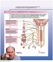
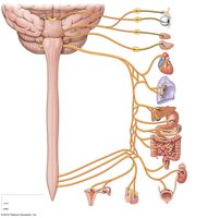
Sympathetic Division
Organization and Anatomy
The sympathetic division consists of preganglionic neurons (T1–L2), ganglia near the vertebral column, and specialized neurons in the suprarenal (adrenal) gland.
Sympathetic Chain Ganglia: Paravertebral ganglia controlling effectors in the body wall, head, neck, limbs, and thoracic cavity.
Collateral Ganglia: Prevertebral ganglia innervating abdominopelvic organs.
Suprarenal Medullae: Modified ganglia releasing hormones (epinephrine and norepinephrine).
Sympathetic Chain Ganglia
Clusters of ganglia parallel to the spinal cord, with specific numbers in each region:
3 cervical
11–12 thoracic
2–5 lumbar
4–5 sacral
1 coccygeal
Collateral Ganglia and Splanchnic Nerves
Splanchnic nerves are bundles of preganglionic fibers converging on collateral ganglia, which innervate visceral organs in the abdominopelvic cavity.
Celiac ganglion: Stomach, duodenum, liver, gallbladder, pancreas, spleen, kidney
Superior mesenteric ganglion: Small intestine, initial large intestine
Inferior mesenteric ganglion: Terminal large intestine, kidney, bladder, sex organs

Suprarenal Medullae
Preganglionic fibers innervate the adrenal medulla, causing release of epinephrine (adrenaline) and norepinephrine into the bloodstream, affecting metabolic activity throughout the body.

Effects of Sympathetic Stimulation
Increased alertness and energy
Enhanced cardiovascular and respiratory activity
Mobilization of energy reserves
Dilation of pupils and respiratory tubes
Stimulation of sweat glands and arrector pili muscles
Neurotransmitter Release and Receptors
Sympathetic ganglion fibers release acetylcholine (ACh) at synapses with ganglionic neurons (cholinergic synapses). Most postganglionic fibers release norepinephrine (adrenergic terminals), but some release ACh.
Alpha receptors: Respond to epinephrine and norepinephrine
Beta receptors: Respond to epinephrine
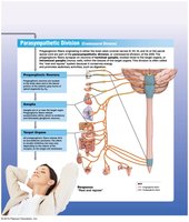
Parasympathetic Division
Organization and Anatomy
Preganglionic neurons are located in the brainstem and sacral segments (S2–S4). Postganglionic neurons are near or within the target organ.
Cranial Nerves: CN III, VII, IX, X
Sacral Nerves: Pelvic nerves
General Functions
Relaxation and energy absorption
Food processing and nutrient absorption
Pupil constriction
Secretion of digestive enzymes and hormones
Increased smooth muscle activity in the digestive system
Stimulation of defecation and urination
Reduced heart rate
Sexual arousal
Neurotransmitter Release and Receptors
All parasympathetic fibers release ACh. Effects are localized and brief due to rapid breakdown by acetylcholinesterase.
Nicotinic receptors: Found on ganglionic neurons; always excitatory
Muscarinic receptors: Found on neuroeffector junctions; can be excitatory or inhibitory
Relationships Between Sympathetic and Parasympathetic Divisions
Dual Innervation
Most vital organs receive input from both divisions, often with opposing (antagonistic) effects. This dual innervation allows fine control of organ function.
Head: Parasympathetic fibers accompany sympathetic fibers
Thoracic/Abdominopelvic: Fibers mingle in plexuses (cardiac, pulmonary, esophageal, celiac, mesenteric, hypogastric)

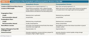
Comparison Table: Sympathetic vs. Parasympathetic Divisions
The following table summarizes key differences:
Characteristic | Sympathetic Division | Parasympathetic Division |
|---|---|---|
Location of CNS Visceral Motor Neurons | Lateral gray horns of spinal segments T1–L2 | Brain stem and sacral segments S2–S4 |
Location of PNS Ganglia | Near spinal cord (sympathetic chain, collateral, suprarenal medullae) | Near or within target organs (terminal/intramural ganglia) |
Preganglionic Fibers | Relatively short, myelinated | Relatively long, myelinated |
Neurotransmitter Released | Acetylcholine | Acetylcholine |
Postganglionic Fibers | Relatively long, unmyelinated | Relatively short, unmyelinated |
Neurotransmitter Junction | Varicosities release norepinephrine (most), some release ACh | Neuroeffector junctions release ACh |
Degree of Divergence from CNS | Extensive (1:32) | Minimal (1:6) |
General Functions | Prepare for emergencies, increase alertness, energy | Promote relaxation, stable energy storage |
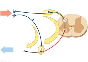
Visceral Reflexes
Types of Visceral Reflexes
Visceral reflexes provide autonomic motor responses and are polysynaptic. They can be classified as:
Long Reflexes: Sensory neurons send signals to the CNS, which processes and sends motor commands to visceral organs.
Short Reflexes: Sensory impulses go directly to ganglionic neurons, bypassing the CNS; motor commands are distributed by postganglionic fibers.
Summary
The autonomic nervous system is essential for maintaining internal balance and responding to environmental changes. Its sympathetic and parasympathetic divisions work together, often antagonistically, to regulate organ function. Understanding the structure, neurotransmitters, and reflex pathways of the ANS is crucial for comprehending human physiology.
