 Back
BackAxial and Appendicular Skeleton: Key Bones and Landmarks
Study Guide - Smart Notes
 Tailored notes based on your materials, expanded with key definitions, examples, and context.
Axial Skeleton
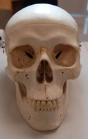
Skull: Major Bones and Foramina
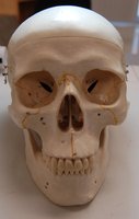
The skull is a complex structure composed of cranial and facial bones, protecting the brain and supporting facial structures. Key features include foramina for nerves and blood vessels, and sutures joining the bones.
Frontal Bone: Forms the forehead and superior part of the orbit.
Parietal Bones: Paired bones forming the superior and lateral aspects of the cranium.
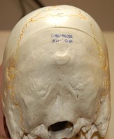
Occipital Bone: Forms the posterior and base of the skull; contains the foramen magnum for the spinal cord.
Temporal Bones: Paired bones at the sides and base of the skull, housing the structures of the ears.
Sphenoid Bone: Forms part of the cranial floor and unites the cranial and facial bones.
Ethmoid Bone: Contributes to the medial wall of the orbit, nasal septum, and nasal cavity.
Maxilla: Upper jaw bone, forms part of the orbit, nasal cavity, and hard palate.
Zygomatic Bones: Cheekbones, form part of the orbit.
Mandible: Lower jaw bone, the only movable skull bone.
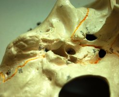
Foramina: Openings such as the supraorbital foramen, infraorbital foramen, mental foramen, and foramen magnum allow passage of nerves and vessels.
Skull Sutures and Landmarks
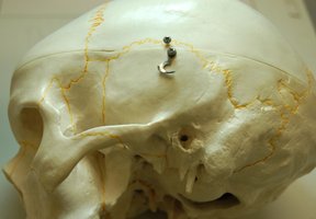
Sutures are immovable joints connecting the bones of the skull. Important landmarks include the coronal, sagittal, lambdoid, and squamous sutures.
Coronal Suture: Between the frontal and parietal bones.
Lambdoid Suture: Between the parietal and occipital bones.
Squamous Suture: Between the parietal and temporal bones.
Other Landmarks: External occipital protuberance, mastoid process, styloid process.
Mandible and Maxilla
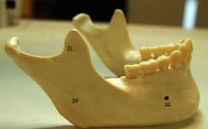
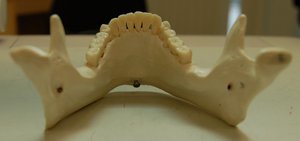
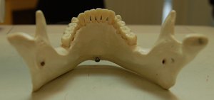
The mandible is the largest and strongest facial bone, forming the lower jaw. The maxilla forms the upper jaw and part of the hard palate.
Mandibular Foramen: Passage for the inferior alveolar nerve.
Mental Foramen: Passage for nerves and vessels to the chin.
Alveolar Margin: Ridge containing the tooth sockets.
Vertebral Column
Vertebrae: Structure and Classification
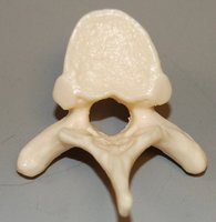
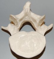
The vertebral column consists of 33 vertebrae, classified into cervical, thoracic, lumbar, sacral, and coccygeal regions. Each vertebra has a body, vertebral foramen, and processes for muscle and ligament attachment.
Cervical Vertebrae (C1–C7): Smallest, with transverse foramina. C1 (atlas) and C2 (axis) are specialized for head movement.
Thoracic Vertebrae (T1–T12): Articulate with ribs, have long spinous processes.
Lumbar Vertebrae (L1–L5): Largest, support most body weight.
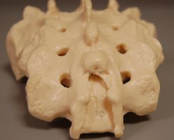
Sacrum: Five fused vertebrae forming the posterior pelvis.
Coccyx: Four fused vertebrae forming the tailbone.
Thoracic Cage
Sternum and Ribs
The thoracic cage protects vital organs and supports the shoulder girdle. It consists of the sternum, ribs, and thoracic vertebrae.
Sternum: Composed of the manubrium, body, and xiphoid process.
Ribs: Twelve pairs, classified as true (1–7), false (8–12), and floating (11–12).
Appendicular Skeleton
Upper Limb: Humerus
The humerus is the longest bone of the upper limb, articulating proximally with the scapula and distally with the radius and ulna.
Head: Articulates with the glenoid fossa of the scapula.
Greater and Lesser Tubercles: Sites for muscle attachment.
Deltoid Tuberosity: Attachment for the deltoid muscle.
Medial and Lateral Epicondyles: Attachment for forearm muscles.
Olecranon Fossa: Receives the olecranon of the ulna during elbow extension.
Lower Limb: Femur, Tibia, and Fibula
The femur is the longest and strongest bone in the body, articulating with the pelvis and tibia. The tibia and fibula form the bones of the lower leg.
Femur: Features include the head, neck, greater and lesser trochanters, and linea aspera.
Tibia: Medial bone of the leg, with the tibial tuberosity and medial malleolus.
Fibula: Lateral bone of the leg, with the head and lateral malleolus.
Foot: Tarsals, Metatarsals, and Phalanges
The foot consists of tarsal bones (including the calcaneus and talus), metatarsals, and phalanges, providing support and mobility.
Tarsals: Seven bones including the calcaneus (heel) and talus (ankle joint).
Metatarsals: Five long bones of the foot.
Phalanges: Fourteen bones forming the toes.
Summary Table: Major Bones and Landmarks
Bone | Key Landmarks | Function/Significance |
|---|---|---|
Skull (Cranium & Face) | Foramina, Sutures, Processes | Protects brain, supports facial structures |
Mandible | Mental foramen, alveolar margin | Lower jaw, mastication |
Vertebrae | Body, foramen, processes | Supports body, protects spinal cord |
Sternum & Ribs | Manubrium, xiphoid, costal cartilages | Protects thoracic organs |
Humerus | Head, tubercles, epicondyles | Upper limb movement |
Femur | Head, trochanters, linea aspera | Weight bearing, lower limb movement |
Tibia & Fibula | Tuberosity, malleoli | Support and movement of leg |
Foot Bones | Calcaneus, talus, metatarsals | Support, balance, locomotion |
