 Back
BackBlood: Structure, Function, and Clinical Relevance (Chapter 19 Study Notes)
Study Guide - Smart Notes
 Tailored notes based on your materials, expanded with key definitions, examples, and context.
Blood: Structure, Function, and Clinical Relevance
Overview of Blood
Blood is a specialized liquid connective tissue that interacts with all other body systems. It is essential for transportation, regulation, and protection within the human body.
Transportation: Delivers gases (O2, CO2), nutrients, and metabolic wastes.
Regulation: Maintains pH, temperature, and water content of cells.
Protection: Provides immune defense and enables clotting to prevent blood loss.
Blood is more viscous than water and ranges in color from bright scarlet (high O2) to brick red (low O2). It has a pH of 7.35–7.45 and constitutes about 8% of body weight (5–6 L in males, 4–5 L in females).
Components of Blood
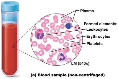
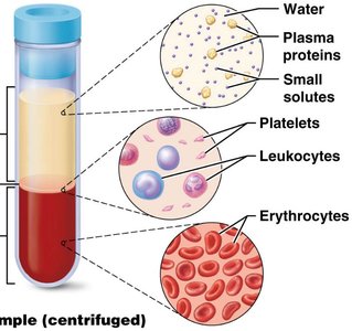
Blood consists of cells (formed elements) and plasma (the liquid matrix).
Red Blood Cells (RBCs, Erythrocytes): ~44% of blood volume
White Blood Cells (WBCs) & Platelets: ~1% of blood volume ("buffy coat")
Plasma: ~55% of blood volume; 90% water, 9% proteins (albumins, globulins, fibrinogen, regulatory proteins), 1% solutes (nutrients, wastes, gases)
Red Blood Cells (Erythrocytes)
Structure and Function
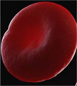
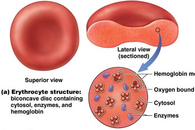
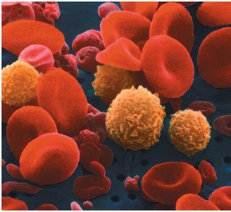
Red blood cells are anucleate, biconcave discs that maximize surface area for gas exchange and lack organelles to provide more space for hemoglobin (Hb).
Normal count: ~5.4 million/μL of blood
Functional lifespan: up to 120 days
Primary function: Transport O2 and CO2
Hemoglobin
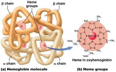
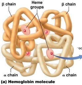
Hemoglobin is the oxygen-carrying protein in RBCs. Each molecule consists of four polypeptide chains (globins), each with a heme group containing iron that binds O2.
Each Hb molecule binds up to 4 O2 molecules.
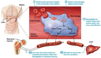
Hb is recycled after RBCs are phagocytosed in the spleen and liver.
Life Cycle and Recycling of RBCs
Old RBCs are removed by macrophages in the spleen and liver.
Iron and amino acids are recycled; heme is converted to bilirubin and excreted.
Hematopoiesis: Formation of Blood Cells
Hematopoietic Stem Cells (HSCs)
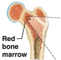
Hematopoiesis is the process of blood cell formation, occurring in red bone marrow. Hematopoietic stem cells can differentiate into any type of blood cell.
Myeloid cell line: Produces RBCs, platelets, and most WBCs
Lymphoid cell line: Produces lymphocytes
Erythropoiesis
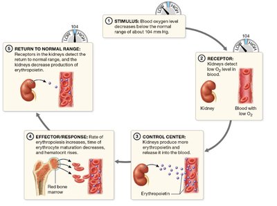
Erythropoiesis is the production of RBCs, regulated by erythropoietin (EPO) from the kidneys in response to hypoxia (low O2 levels).
Requires iron, amino acids, and B vitamins.
Balance between RBC production and destruction is critical for homeostasis.
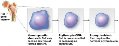
Stages of Erythropoiesis
Stage | Description |
|---|---|
Hematopoietic stem cell | Multipotent cell in red bone marrow |
Erythrocyte-CFU | Committed to becoming an erythrocyte |
Proerythroblast | Requires erythropoietin for further development |
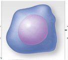
Early erythroblast | Rapid hemoglobin synthesis |
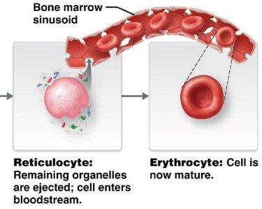
Late erythroblast | Nucleus ejected |
Reticulocyte | Enters bloodstream, matures into erythrocyte |
Disorders of Erythrocytes
Anemia
Anemia is a condition characterized by reduced oxygen-carrying capacity of blood due to low hemoglobin or RBC count.
Iron-deficiency anemia: Most common; insufficient iron for Hb production.
Pernicious anemia: Low RBC production due to vitamin B12 deficiency.
Sickle cell disease: Abnormal Hb causes RBCs to sickle and rupture, leading to severe anemia in homozygous individuals.
White Blood Cells (Leukocytes)
Types and Functions
Leukocytes are nucleated cells involved in immune defense. They leave circulation to combat infection and inflammation.
Granulocytes:
Neutrophils: Phagocytic, multi-lobed nucleus
Eosinophils: Combat parasitic worms, bi-lobed nucleus
Basophils: Release histamine, S-shaped nucleus obscured by granules
Agranulocytes:
Monocytes: U-shaped nucleus, become macrophages
Lymphocytes: B cells (antibody production), T cells (cell-mediated immunity)
Leukopoiesis
Leukopoiesis is the formation of WBCs from hematopoietic stem cells. The lymphoid line produces lymphocytes, while the myeloid line produces other WBCs and RBCs.
Bone marrow and cord blood transplants can treat blood disorders such as anemia and leukemia.
Platelets (Thrombocytes)
Structure and Function
Platelets are small, anucleate cell fragments derived from megakaryocytes. They are essential for blood clotting and are recycled by the spleen and liver after 7–10 days.
Normal count: 250,000–400,000/μL
Contain proteins that promote clotting
Hemostasis and Blood Clotting
Hemostasis
Hemostasis is the process of stopping bleeding through a series of steps:
Vascular spasm: Immediate vasoconstriction of damaged vessel
Platelet plug formation: Platelets adhere to exposed collagen and each other
Coagulation: Formation of a stable clot via activation of clotting factors and fibrin strands
Clotting Cascade
The clotting cascade involves a series of enzymatic reactions:
Formation of prothrombinase (via extrinsic or intrinsic pathway)
Conversion of prothrombin to thrombin
Thrombin converts fibrinogen to fibrin, forming the clot
After healing, clots are retracted and dissolved.
Blood Groups and Transfusion Compatibility
Antigens and Antibodies
Blood groups are defined by the presence of antigens on RBC membranes and antibodies in plasma. Agglutination (clumping) occurs when incompatible antigens and antibodies bind, potentially causing hemolysis and death.
Type O: Universal donor (no antigens on RBCs)
Type AB: Universal recipient (no antibodies in plasma)
Cross-matching with antisera identifies blood type for safe transfusion.
Rh Factor and Hemolytic Disease of the Newborn
Rh-: RBCs lack Rh antigen
Rh+: RBCs have Rh antigen
Hemolytic disease of the newborn can occur if an Rh- mother carries an Rh+ fetus, leading to maternal antibody production and fetal RBC destruction in subsequent pregnancies.
Summary Table: Main Components of Blood
Component | Percentage of Blood | Main Function |
|---|---|---|
Plasma | ~55% | Transport of nutrients, wastes, proteins, hormones |
Red Blood Cells | ~44% | Oxygen and carbon dioxide transport |
White Blood Cells | ~1% | Immune defense |
Platelets | ~1% | Blood clotting |
Additional info: These notes integrate and expand upon the provided lecture slides, filling in academic context for clarity and exam preparation.
