 Back
BackBrain Structure, Protection, and Major Functional Systems
Study Guide - Smart Notes
 Tailored notes based on your materials, expanded with key definitions, examples, and context.
Brain Terminology & Major Regions
Cerebrum of the Brain
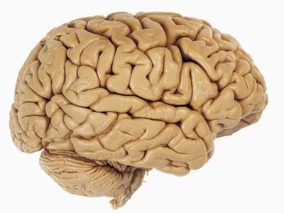
The cerebrum is the largest part of the brain and is responsible for higher brain functions such as thought, memory, and voluntary movement. It is divided into two hemispheres (right and left), each covered by the cerebral cortex, a layer of gray matter. The surface of the cerebrum is marked by gyri (elevated ridges), sulci (shallow depressions), and fissures (deeper grooves), all of which increase the surface area for neural processing.
Major Parts of the Brain
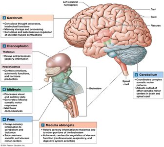
The brain is organized into four main regions, each with specialized functions:
Cerebrum: Responsible for conscious thought, memory, and voluntary muscle contractions.
Cerebellum: Coordinates complex somatic motor patterns and maintains posture and balance.
Diencephalon: Contains the thalamus (relay and processing center for sensory information) and hypothalamus (centers for emotions, autonomic functions, and hormone production).
Brainstem: Includes the midbrain (processes visual/auditory data, maintains consciousness), pons (relays sensory information, controls respiration), and medulla oblongata (relays sensory information, regulates autonomic functions).
Ventricular System
Ventricles and Connecting Passageways
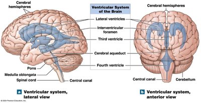
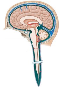
The ventricular system consists of interconnected cavities within the brain that produce and circulate cerebrospinal fluid (CSF). The main ventricles are:
Lateral ventricles: One in each cerebral hemisphere.
Third ventricle: Located in the diencephalon.
Fourth ventricle: Located between the pons and cerebellum, extending into the medulla oblongata.
Passageways include the interventricular foramina (connecting lateral ventricles to the third ventricle) and the cerebral aqueduct (connecting the third and fourth ventricles). The fourth ventricle narrows to become the central canal of the spinal cord.
Cranial Meninges
Dura Mater
The dura mater is the outermost meningeal layer, divided into two sublayers:
Periosteal cranial dura: Fused to the periosteum of cranial bones.
Meningeal cranial dura: Inner layer, can form dural folds and contains venous sinuses for blood drainage.
The subdural space lies beneath the dura mater and is normally empty except in cases of disease or trauma.
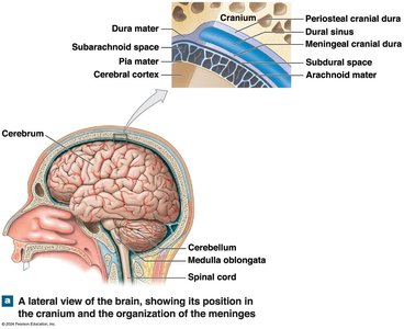
Arachnoid Mater
The arachnoid mater is the middle meningeal layer, resembling a spider web. The subarachnoid space beneath it contains a delicate network of collagen and elastic fibers (arachnoid trabeculae) and is filled with CSF.
Pia Mater
The pia mater is the innermost layer, anchored to the brain's surface by astrocytes and surrounding cerebral blood vessels that penetrate the brain.
Cerebrospinal Fluid (CSF)
Functions of CSF
Cerebrospinal fluid serves several critical functions:
Cushions the brain against physical trauma
Provides buoyancy and support, reducing the effective weight of the brain
Transports nutrients (glucose, amino acids, vitamins, minerals), gases (O2, CO2), and wastes (CO2, lactic acid, ammonia, urea)
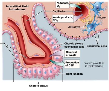
Creation and Circulation of CSF
CSF is produced by the choroid plexus in the ventricles, consisting of specialized ependymal cells and permeable capillaries. It is produced in the third and fourth ventricles and extends into the lateral ventricles. CSF flows through the ventricular system, enters the subarachnoid space via openings in the fourth ventricle, circulates around the brain and spinal cord, and is reabsorbed through arachnoid granulations in the superior sagittal sinus.
CSF Production & Circulation | Details |
|---|---|
Daily Production | ~500 mL |
Total Volume | ~150 mL |
Recycling Rate | Every ~8 hours |
Clinical Note | Hydrocephalus: CSF buildup due to blockage or reabsorption issues |
Blood Supply & Blood-Brain Barrier (BBB)
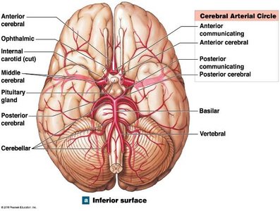
Blood Supply
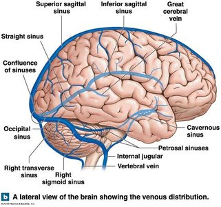
The brain requires a rich blood supply to meet its high oxygen and energy demands. Major arteries include the internal carotid arteries and vertebral arteries, while veins drain into dural sinuses, with the largest being the superior sagittal sinus. All sinuses ultimately drain into the internal jugular vein.
Blood Supply Disorders
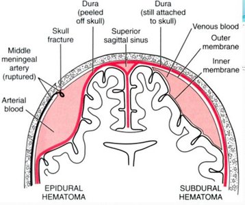
Epidural hemorrhage: Blood accumulates between the dura mater and cranium due to damaged blood vessels.
Subdural hemorrhage: Blood pools between the dura mater and arachnoid mater.
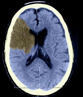
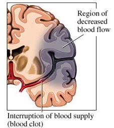
Cerebrovascular accident (CVA, or stroke): Blood supply to a brain region is cut off, causing loss of function in that area.
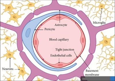
Blood-Brain Barrier (BBB)
The blood-brain barrier is formed by astrocytes wrapping around blood vessels, creating a selective barrier that tightly regulates the interstitial fluid around neurons. Damage to astrocytes can compromise the BBB. A special case is the blood-CSF barrier at the choroid plexus.
Medical Treatments and the BBB
The BBB restricts the passage of many substances, making drug delivery to the brain challenging. Only lipid-soluble substances can easily cross; others may require direct injection into the CSF (e.g., via lumbar puncture). This is an active area of pharmaceutical research.
Antidepressants and the BBB
Selective Serotonin Reuptake Inhibitors (SSRIs) are antidepressants that are typically lipid-soluble, allowing them to cross the BBB. Once in the brain, they inhibit serotonin reuptake, increasing its concentration at synapses and potentially alleviating depression symptoms.
Brainstem & Cerebellum
Medulla Oblongata
The medulla oblongata is the most inferior brain region, containing nuclei that control visceral functions (e.g., heart rate, breathing), relay sensory and motor information, and serve as reflex centers. The reticular formation within the medulla is crucial for maintaining consciousness and alertness.
Pons
The pons links the cerebellum to other brain regions and contains nuclei for cranial nerves V-VIII. It plays a role in respiration, relays messages to/from the cerebellum, and allows communication between the cerebellar hemispheres.
Midbrain
The midbrain regulates visual and auditory reflexes, controls alertness, and relays motor commands. Key structures include the tectum (superior and inferior colliculi), cerebral peduncles, substantia nigra (dopaminergic neurons), red nucleus, and the reticular activating system (RAS).
Cerebellum
The cerebellum adjusts postural muscles to maintain balance and equilibrium and fine-tunes learned movements. It receives extensive sensory input and adjusts motor commands based on proprioceptive, visual, touch, balance, and auditory information.
Diencephalon
Pineal Gland
The pineal gland produces melatonin, a hormone important for regulating day-night cycles and overall health.
Thalamus
The thalamus is the relay point for all ascending sensory information (except olfactory), filtering and passing selected information to the cerebral cortex. It consists of multiple nuclei and coordinates with the basal nuclei and limbic system. The left and right thalami are connected by the interthalamic adhesion.
Hypothalamus
The hypothalamus contains many nuclei with diverse functions, including:
Regulation of autonomic functions (heart rate, blood pressure, respiration, digestion)
Production of regulatory hormones (controls pituitary gland)
Secretion of ADH and oxytocin
Control of emotions and behavioral drives (feeding, fighting, fleeing, reproduction)
Coordination of voluntary and autonomic functions
Regulation of body temperature and circadian rhythms
Limbic System
Functions and Major Structures
The limbic system is a functional grouping involved in motivation, emotion, and memory. It establishes emotional states, links intellectual and unconscious functions, and facilitates memory storage and retrieval (episodic, factual, and emotional).
Hippocampus: Important for learning and long-term memory storage/retrieval.
Amygdala: Involved in aggression, fear, anxiety, and linking emotions with memories.
Cingulate gyrus: Links behavioral outcomes to emotion.
