 Back
BackCell Biology: Structure and Function of the Cell
Study Guide - Smart Notes
 Tailored notes based on your materials, expanded with key definitions, examples, and context.
Cell Biology
Introduction to the Cell
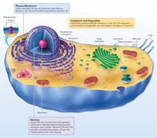
The cell is the fundamental unit of life, responsible for carrying out all essential biological processes. Each cell is bounded by a plasma membrane and contains cytoplasm, which houses the nucleus and various organelles that perform specialized functions.
Plasma (cell) membrane: The outer boundary that regulates interactions with the external environment.
Nucleus: Directs cellular activities and contains genetic material.
Cytoplasm: The region between the plasma membrane and nucleus, containing organelles.
3.1 Functions of the Cell
Characteristic Functions
Metabolism and Energy Use: All chemical reactions within the cell, including energy transfer and heat production.
Synthesis of Molecules: Cells synthesize proteins, nucleic acids, and lipids specific to their function.
Communication: Cells send and receive electrical and chemical signals for coordination.
Reproduction and Inheritance: Each cell contains DNA, which determines its structure and function; some cells (gametes) transmit genetic information to the next generation.
3.2 How We See Cells
Microscopy
Cells are too small to be seen with the naked eye and require microscopes for visualization.
Light Microscope: Resolution ~0.1 µm; used for tissues and cells, often with stains.
Electron Microscope: Resolution ~0.1 nm; includes SEM (surface features) and TEM (internal structures).
Atomic Force Microscope (AFM): Uses a probe to scan the sample, revealing surface topography at very high resolution.
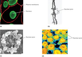
3.3 Plasma Membrane
Structure and Function
The plasma membrane separates intracellular from extracellular environments, supports cell contents, attaches to other cells or the extracellular matrix, enables recognition and communication, and regulates the movement of substances.
Membrane Potential: An electrical charge difference across the membrane due to regulated ion movement; more positive ions outside, more negative ions and proteins inside.
3.4 Membrane Lipids
Phospholipids and Cholesterol
The plasma membrane is primarily composed of a phospholipid bilayer and cholesterol, with small amounts of carbohydrates.
Phospholipids: Form a bilayer with hydrophilic heads facing water and hydrophobic tails facing inward.
Cholesterol: Interspersed among phospholipids, stabilizing membrane fluidity.
Glycocalyx: Carbohydrate-rich area on the cell surface, important for cell recognition.
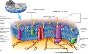
3.5 Membrane Proteins
Types and Functions
Integral Proteins: Span the membrane; may form channels or act as carriers.
Peripheral Proteins: Attached to membrane surfaces; function as markers, attachment sites, enzymes, or receptors.
Marker Molecules: Glycoproteins/glycolipids for cell recognition (e.g., distinguishing self from foreign cells).
Attachment Proteins: Cadherins (cell-cell attachment), integrins (cell-matrix attachment).
Transport Proteins: Channels, carriers, and ATP-powered pumps for selective transport.
Receptor Proteins: Bind specific ligands to trigger cellular responses.
Enzymes: Catalyze reactions at the membrane surface.
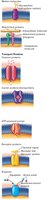
3.6 Movement through the Plasma Membrane
Membrane Transport Mechanisms
The plasma membrane is selectively permeable, allowing only certain substances to pass. Transport mechanisms include:
Passive Transport: No ATP required; substances move down their concentration gradient (diffusion, osmosis, facilitated diffusion).
Active Transport: Requires ATP; substances move against their concentration gradient (active transport, secondary active transport).
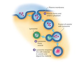
Vesicular Transport: Movement via vesicles (endocytosis, exocytosis, transcytosis).
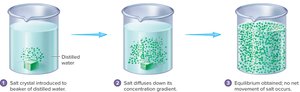
Diffusion
Net movement of solutes from high to low concentration until equilibrium is reached.
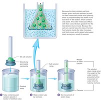
Osmosis
Diffusion of water across a selectively permeable membrane from low to high solute concentration. Osmotic pressure is the force required to prevent water movement.
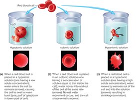
Effects of Tonicity on Cells
Isotonic: No net water movement; cell remains normal.
Hypertonic: Water leaves the cell; cell shrinks (crenation).
Hypotonic: Water enters the cell; cell swells or bursts (lysis).
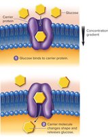
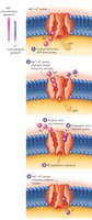
Facilitated Diffusion
Carrier or channel proteins help move large or charged molecules across the membrane without ATP.
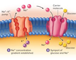
Active Transport
ATP is used to move substances against their concentration gradient. The sodium-potassium pump is a key example, moving 3 Na+ out and 2 K+ into the cell per ATP hydrolyzed.
Secondary Active Transport
Uses the energy from the concentration gradient of one substance (e.g., Na+) to transport another substance (e.g., glucose) against its gradient.
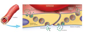
Vesicular Transport
Endocytosis: Uptake of materials via vesicle formation (phagocytosis for solids, pinocytosis for liquids).
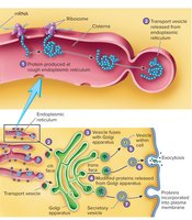
Exocytosis: Release of substances from the cell via vesicle fusion with the plasma membrane.
Transcytosis: Movement through a cell by endocytosis and exocytosis.
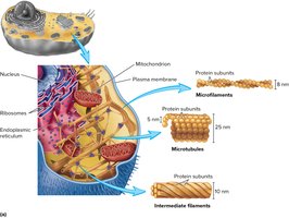
3.7 Cytoplasm
Components
Cytosol: Fluid portion containing dissolved molecules and ions.
Cytoskeleton: Network of protein filaments (microtubules, actin filaments, intermediate filaments) providing support, shape, and movement.
Cytoplasmic Inclusions: Aggregates of chemicals such as lipid droplets, glycogen, and pigments.
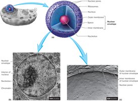
3.8 The Nucleus and Cytoplasmic Organelles
Nucleus
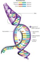
The nucleus is a large, membrane-bound organelle containing DNA. It is surrounded by a double membrane (nuclear envelope) with nuclear pores for molecular exchange. The nucleolus within the nucleus is the site of ribosome production.
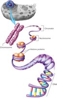
Chromosome Structure
DNA associates with histone proteins to form chromatin, which condenses into chromosomes during cell division. Nucleosomes are the basic units of chromatin structure.
Ribosomes
Sites of protein synthesis, composed of rRNA and proteins. Free ribosomes synthesize intracellular proteins, while those attached to the rough ER produce proteins for secretion or membrane insertion.
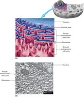
Endoplasmic Reticulum (ER)
Rough ER: Studded with ribosomes; synthesizes and modifies proteins.
Smooth ER: Lacks ribosomes; synthesizes lipids, detoxifies chemicals, stores calcium ions.
Golgi Apparatus
Stack of flattened membranes that modifies, packages, and distributes proteins and lipids. Forms vesicles for secretion, membrane insertion, or lysosome formation.
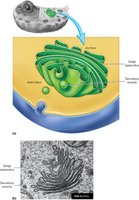
Lysosomes and Peroxisomes
Lysosomes: Contain hydrolytic enzymes for digesting cellular debris and foreign material.
Peroxisomes: Contain enzymes for breaking down fatty acids and amino acids; detoxify hydrogen peroxide.
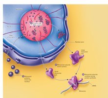
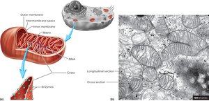
Mitochondria
Major site of ATP synthesis. Contains an outer and highly folded inner membrane (cristae) and its own DNA. The matrix contains enzymes for the citric acid cycle.
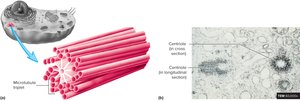
Centrioles and Spindle Fibers
Centrioles are involved in microtubule formation and cell division. Spindle fibers organize and separate chromosomes during mitosis.
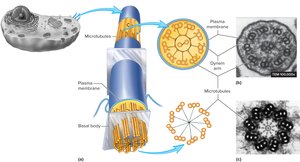
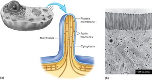
Cilia, Flagella, and Microvilli
Cilia: Move materials over cell surfaces (e.g., mucus in the respiratory tract).
Flagella: Propel sperm cells.
Microvilli: Increase surface area for absorption; supported by actin filaments.
3.9 Genes and Gene Expression
Gene Expression
Genes are DNA segments that code for RNA or proteins. Gene expression involves transcription (DNA to mRNA) and translation (mRNA to protein).
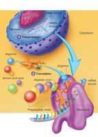
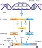
Transcription
RNA polymerase synthesizes mRNA from DNA template. Introns are removed from pre-mRNA, and exons are spliced together. A 7-methylguanosine cap and poly-A tail are added for stability and export.
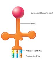
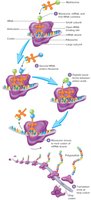
Translation
Occurs on ribosomes; tRNA brings amino acids matching mRNA codons, and rRNA catalyzes peptide bond formation. The process continues until a stop codon is reached.
3.10 Cell Cycle
Phases of the Cell Cycle
Interphase: Cell growth, DNA replication, and preparation for division (G1, S, G2 phases).
Mitosis: Division of the nucleus (prophase, metaphase, anaphase, telophase).
Cytokinesis: Division of the cytoplasm, resulting in two daughter cells.
DNA replication ensures genetic continuity. Chromatin condenses into chromosomes, which are separated during mitosis.
Summary Table: Membrane Transport Mechanisms
Mechanism | Energy Required | Direction | Example |
|---|---|---|---|
Diffusion | No | High to Low | O2, CO2 |
Osmosis | No | Water: Low to High Solute | Water movement |
Facilitated Diffusion | No | High to Low | Glucose transport |
Active Transport | Yes (ATP) | Low to High | Na+/K+ pump |
Secondary Active Transport | Indirect (ATP) | Low to High (for one substance) | Na+-glucose symport |
Vesicular Transport | Yes (ATP) | Varies | Endocytosis, exocytosis |
