 Back
BackCells and Mitosis: Structure, Function, and Division
Study Guide - Smart Notes
 Tailored notes based on your materials, expanded with key definitions, examples, and context.
Cells: The Structural Units of Life
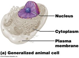
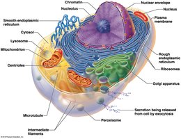
Anatomy of a Generalized Cell
Cells are the basic structural and functional units of all living organisms. Each cell typically consists of three main regions: the plasma membrane, nucleus, and cytoplasm. These components work together to maintain cellular integrity and function.
Plasma Membrane: Serves as a barrier, separating cell contents from the external environment.
Nucleus: Acts as the control center, housing genetic material (DNA).
Cytoplasm: The site of most cellular activities, containing organelles suspended in cytosol.
The Plasma Membrane
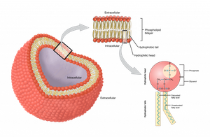
Structure and Function
The plasma membrane is a selectively permeable barrier composed of a double layer of phospholipids arranged "tail to tail," with cholesterol and proteins interspersed. This structure is described by the fluid mosaic model.
Phospholipid Bilayer: Provides fluidity and flexibility to the membrane.
Proteins: Serve as channels, receptors, and enzymes.
Cholesterol: Stabilizes membrane structure.
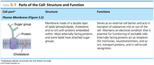
Cell Part | Structure | Functions |
|---|---|---|
Plasma Membrane | Double layer of lipids with proteins, cholesterol, and some sugars | Acts as a barrier, controls entry/exit, cell recognition, and communication |
The Nucleus
Structure and Function
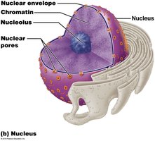
The nucleus is the cell's control center, containing DNA necessary for protein synthesis and cell reproduction. It consists of three main regions:
Nuclear Envelope: Double membrane with pores for material exchange.
Nucleolus: Site of ribosome synthesis.
Chromatin: DNA and protein complex that condenses into chromosomes during cell division.
The Cytoplasm
Major Organelles and Their Functions
The cytoplasm is the cellular material outside the nucleus and inside the plasma membrane. It contains various organelles, each with specialized functions:
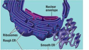
Endoplasmic Reticulum (ER): Network of membranes continuous with the nuclear envelope.
Rough ER: Studded with ribosomes; synthesizes proteins.
Smooth ER: Lacks ribosomes; involved in lipid metabolism and detoxification.
Ribosomes: Sites of protein synthesis; found free in cytoplasm or attached to rough ER.
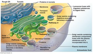
Golgi Apparatus: Modifies, sorts, and packages proteins for secretion or delivery to other organelles.
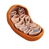
Mitochondria: "Powerhouses" of the cell; generate ATP through cellular respiration.
Centrioles: Direct formation of the mitotic spindle during cell division.
Cell Division
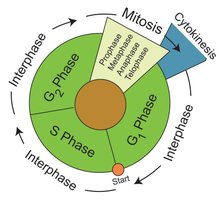
Overview of the Cell Cycle
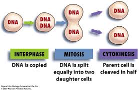
The cell life cycle consists of a series of events from cell formation to division. It includes two major periods: interphase (cell growth and metabolic activity) and cell division (mitosis and cytokinesis).
Interphase: Cell grows, performs normal functions, and duplicates DNA.
Cell Division: Involves mitosis (nuclear division) and cytokinesis (cytoplasmic division).
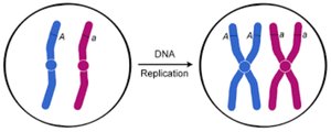
DNA Replication
During interphase, the cell duplicates its genetic material to prepare for division. Each chromosome is copied, resulting in two identical sister chromatids.
Events of Cell Division
Mitosis: Division of the nucleus, producing two genetically identical daughter nuclei.
Cytokinesis: Division of the cytoplasm, resulting in two separate daughter cells.
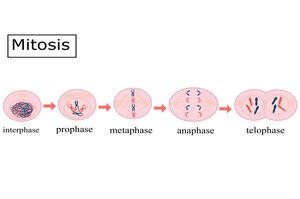
Phases of Mitosis
Prophase: Chromatin condenses into chromosomes; mitotic spindle forms; nuclear envelope breaks down.
Metaphase: Chromosomes align at the cell's equator (metaphase plate).
Anaphase: Sister chromatids separate and move toward opposite poles.
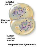
Telophase: Chromosomes uncoil; nuclear envelope reforms; nucleoli reappear.


Cytokinesis
Cytokinesis is the division of the cytoplasm, which usually begins during late telophase. A cleavage furrow forms, pinching the cell into two daughter cells, each with a complete set of organelles and genetic material.
Summary Table: Major Cell Structures and Functions
Organelle | Structure | Function |
|---|---|---|
Plasma Membrane | Phospholipid bilayer with proteins | Selective barrier, cell communication |
Nucleus | Double membrane, nucleolus, chromatin | Genetic control, ribosome synthesis |
Rough ER | Membranous sacs with ribosomes | Protein synthesis |
Smooth ER | Membranous sacs without ribosomes | Lipid metabolism, detoxification |
Golgi Apparatus | Stack of flattened membranes | Protein modification and packaging |
Mitochondria | Double membrane, inner folds (cristae) | ATP production |
Centrioles | Cylindrical structures | Spindle formation in cell division |
Additional info: The above content expands on the provided notes with definitions, examples, and logical academic context to ensure completeness and clarity for college-level anatomy and physiology students.
