 Back
BackCells: The Living Units – Structure, Function, and Membrane Transport
Study Guide - Smart Notes
 Tailored notes based on your materials, expanded with key definitions, examples, and context.
Cell Theory and Cell Diversity
Definition and Importance of Cells
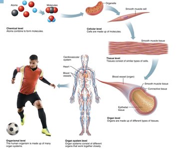
Cells are the fundamental structural and functional units of all living organisms. They are capable of independent replication and contain cytoplasm, a plasma membrane, and various biomolecules such as proteins and nucleic acids. The human body is composed of 50 to 100 trillion cells, each specialized for distinct functions.
Cell Theory: All living things are composed of cells; cells are the smallest units of life; new cells arise only from pre-existing cells.
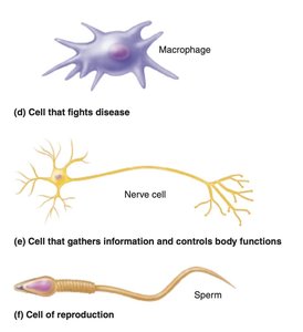
Cell Diversity: Over 250 types of human cells exist, differing in size, shape, and subcellular components. These differences are determined by gene expression and directly influence cell function.
Example: Nerve cells transmit signals, muscle cells contract, and macrophages fight disease.
Structure of a Generalized Cell
Major Regions and Their Functions
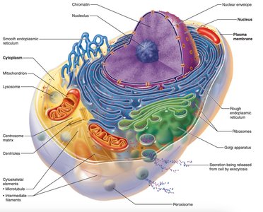
A typical human cell consists of three main regions, each with distinct roles:
Nucleus: The control center containing DNA, regulating gene expression and cell activities such as growth, metabolism, and reproduction.
Cytoplasm: The semifluid interior containing organelles, which perform specialized functions like energy production (mitochondria) and protein synthesis (ribosomes).
Plasma Membrane: The flexible boundary separating intracellular and extracellular environments, controlling the entry and exit of substances.
Extracellular Materials
Composition and Function
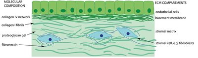
Extracellular materials surround cells, providing structural and biochemical support. They include:
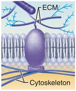
Extracellular Matrix (ECM): A network of proteins (collagen, elastin) and carbohydrates (proteoglycans) that acts as "cell glue," offering strength, elasticity, and hydration.
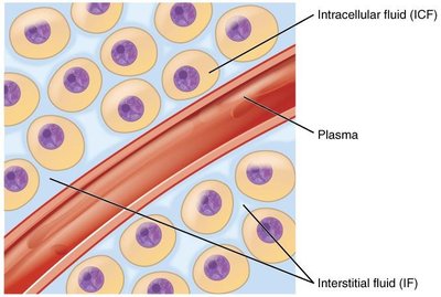
Extracellular Fluid (ECF): The fluid outside cells, including interstitial fluid, blood plasma, and cerebrospinal fluid, which delivers nutrients and removes waste.
Plasma Membrane Structure and Composition
Chemical Components
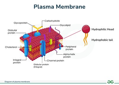
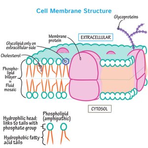
The plasma membrane separates the intracellular fluid from the extracellular fluid and is composed of:
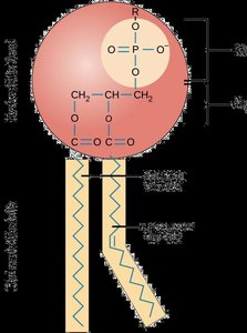
Lipids: Phospholipids (form a bilayer with hydrophilic heads and hydrophobic tails), cholesterol (provides stability and fluidity), glycolipids (cell recognition).
Proteins: Integral (embedded, act as channels/carriers) and peripheral (loosely attached, support membrane function).
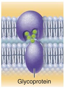
Carbohydrates: Glycoproteins and glycolipids on the outer surface, involved in cell recognition and adhesion.

Membrane Proteins and Their Functions
Types and Roles
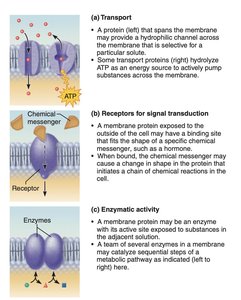
Membrane proteins are essential for various cellular functions:
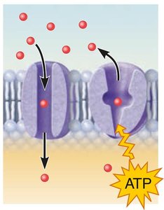
Transport: Channels and carriers regulate the movement of substances across the membrane.
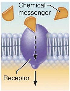
Signal Transduction: Receptors receive and transmit signals from the environment.
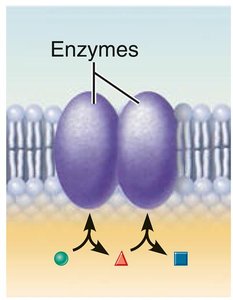
Enzymatic Activity: Enzymes catalyze reactions at the membrane surface.
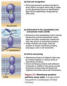
Cell-Cell Recognition: Glycoproteins serve as identification tags for cell recognition.
Cell-to-Cell Joining: Proteins form junctions for tissue integrity.
Attachment: Proteins anchor the cytoskeleton and ECM, maintaining cell shape and facilitating movement.
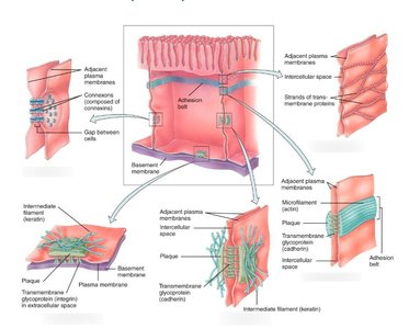
Cell Junctions
Types and Functions
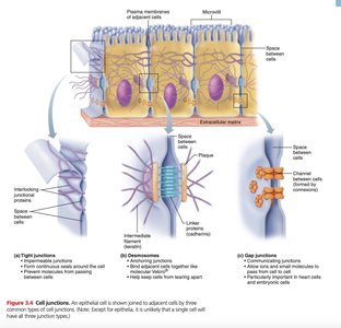
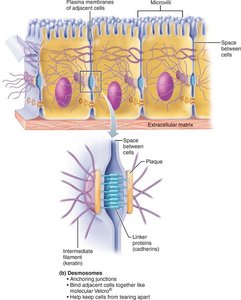
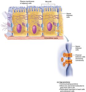
Cell junctions are specialized structures that connect cells, enable communication, and maintain tissue integrity. The three main types are:
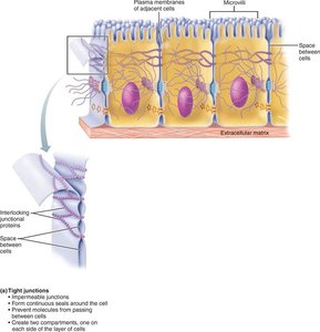
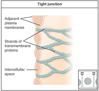
Tight Junctions: Form a seal to prevent leakage of molecules between cells, especially in epithelial tissues.
Desmosomes: Provide strong adhesion through intermediate filaments, abundant in tissues subject to mechanical stress (e.g., skin, heart muscle).
Gap Junctions: Form water-filled channels for direct communication, allowing ions and small molecules to pass between cells, essential for coordinated activity (e.g., heart muscle).
Membrane Transport Mechanisms
Passive Transport
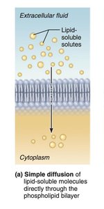
Passive transport allows substances to cross the membrane without energy input. The main types are:
Simple Diffusion: Movement of small, nonpolar molecules (e.g., O2, CO2) directly through the lipid bilayer, from high to low concentration.
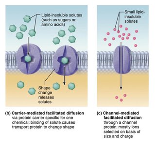
Facilitated Diffusion: Movement of large, polar molecules (e.g., glucose, amino acids) via membrane proteins (channels or carriers), down the concentration gradient.
Osmosis: Movement of water across a selectively permeable membrane, from low solute concentration to high solute concentration, through aquaporins or the lipid bilayer.
Tonicity and Osmosis
Tonicity describes the effect of a solution on cell shape by altering internal water volume:
Isotonic: Equal solute concentrations inside and outside the cell; no net water movement.
Hypertonic: Higher solute concentration outside; water leaves the cell, causing shrinkage.
Hypotonic: Lower solute concentration outside; water enters the cell, causing swelling.
Active Transport
Active transport moves substances against their concentration gradient, requiring energy (ATP):
Primary Active Transport: Direct use of ATP to move substances (e.g., Na+/K+ pump).
Secondary Active Transport: Uses energy stored in ionic gradients created by primary active transport (e.g., glucose cotransport).
Vesicular Transport
Endocytosis and Exocytosis
Vesicular transport moves large substances or amounts across membranes in vesicles:
Endocytosis: Uptake of materials into the cell via vesicle formation. Types include pinocytosis (cell drinking), phagocytosis (cell eating), and receptor-mediated endocytosis (highly specific).
Exocytosis: Release of substances from the cell by vesicle fusion with the plasma membrane.
Membrane Potential
Establishment and Maintenance
Membrane potential is the voltage across the plasma membrane, essential for nerve impulse transmission and muscle contraction. It is established by:
Ion Distribution: Unequal distribution of Na+, K+, and Cl- across the membrane.
Selective Permeability: Ion channels allow specific ions to move, creating electrical gradients.
Sodium-Potassium Pump: Actively transports 3 Na+ out and 2 K+ in, maintaining the electrochemical gradient.
Cell Adhesion Molecules and Membrane Receptors
Cell Interaction and Signaling
Cells interact with their environment through:
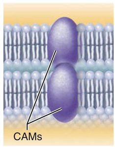
Cell Adhesion Molecules (CAMs): Proteins that enable cell-cell and cell-matrix adhesion, guide cell migration, and stimulate junction formation.
Membrane Receptors: Proteins that bind signaling molecules (ligands) and initiate intracellular responses, including signal transduction, cell growth, differentiation, and immune response.
G Protein-Coupled Receptors (GPCRs): Activate intracellular signaling pathways via G proteins, involved in diverse physiological processes.
Summary Table: Comparing Membrane Transport Mechanisms
Mechanism | Substances Transported | Direction | Energy Required | Example |
|---|---|---|---|---|
Simple Diffusion | Small, nonpolar molecules | High to low concentration | No | O2, CO2 |
Facilitated Diffusion | Large, polar molecules, ions | High to low concentration | No | Glucose, amino acids |
Osmosis | Water | Low solute to high solute | No | Water movement |
Primary Active Transport | Ions | Low to high concentration | Yes (ATP) | Na+/K+ pump |
Secondary Active Transport | Glucose, amino acids | Coupled with ion movement | Indirect (ATP) | Glucose cotransport |
Recap: Key Concepts
Cells are the basic units of life.
Major cell regions: nucleus, cytoplasm, and plasma membrane.
Plasma membrane: selective barrier composed of lipids, proteins, and carbohydrates.
Transport mechanisms: passive (simple diffusion, facilitated diffusion, osmosis) and active transport.
Endocytosis and exocytosis: processes for cellular uptake and release of materials.
Membrane potential: difference in electrical charge across the membrane.
Cell adhesion molecules: enable cell-cell and cell-matrix interactions.
Membrane receptors: mediate cell signaling and communication.
