 Back
BackCentral Nervous System: Brain Structure, Function, and Cranial Nerves
Study Guide - Smart Notes
 Tailored notes based on your materials, expanded with key definitions, examples, and context.
Central Nervous System: Brain Structure, Function, and Cranial Nerves
Introduction to the Central Nervous System (CNS)
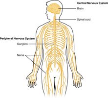
The central nervous system (CNS) consists of the brain and spinal cord, serving as the primary control center for the body. It integrates sensory information and coordinates bodily functions. The peripheral nervous system (PNS) includes all neural elements outside the CNS, such as nerves and ganglia.
Central Nervous System (CNS): Brain and spinal cord, located within the cranial and vertebral cavities.
Peripheral Nervous System (PNS): Nerves and ganglia outside the CNS, connecting the CNS to limbs and organs.
Nervous Tissue Structure
Neurons and Glial Cells
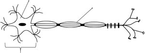
Nervous tissue is composed of two main cell types: neurons and glial cells. Neurons are responsible for transmitting electrical signals, while glial cells provide support and protection.
Neuron: The functional unit of the nervous system, consisting of a cell body (soma), dendrites (receive signals), and an axon (transmits signals).
Glial Cells: Supportive cells that maintain the environment around neurons.
Gray Matter vs. White Matter
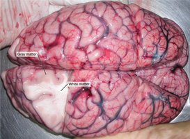
Within the CNS, regions are classified as gray matter or white matter based on their composition and appearance.
Gray Matter: Contains neuron cell bodies, dendrites, and unmyelinated axons. Found in the cerebral cortex and subcortical nuclei.
White Matter: Composed mainly of myelinated axons, which appear white due to the lipid-rich myelin sheath. Found in deeper brain regions and the corpus callosum.
Protective Coverings: The Meninges
Brain Meninges
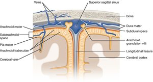
The brain is protected by three connective tissue membranes called meninges, which provide structural support and contain cerebrospinal fluid (CSF).
Dura Mater: The tough, outermost layer, providing a durable protective sheath.
Arachnoid Mater: The middle, web-like layer, with a subarachnoid space filled with CSF.
Pia Mater: The delicate, innermost layer, adhering closely to the brain's surface and following its contours.
Major Brain Regions and Structures
Cerebrum
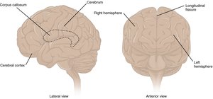
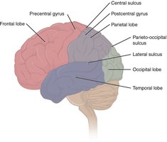
The cerebrum is the largest part of the brain, responsible for higher cognitive functions, voluntary movement, and sensory processing. It is divided into two hemispheres by the longitudinal fissure and further into lobes by sulci and gyri.
Gyri: Elevated ridges on the brain's surface.
Sulci: Shallow grooves between gyri.
Fissures: Deep grooves, such as the longitudinal fissure separating the hemispheres.
Subcortical Gray Matter
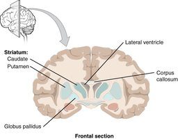
Beneath the cerebral cortex are subcortical nuclei, including the basal nuclei, hippocampus, and amygdala. These structures are involved in movement regulation, memory, and emotion.
Basal Nuclei: Includes the caudate, putamen, and globus pallidus; involved in movement control.
Hippocampus: Essential for long-term memory formation.
Amygdala: Involved in emotional responses.
Diencephalon
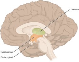
The diencephalon connects the cerebrum to the rest of the nervous system and includes the thalamus and hypothalamus.
Thalamus: Relays sensory and motor signals to the cerebral cortex.
Hypothalamus: Regulates homeostasis, autonomic functions, and the endocrine system.
Brain Stem
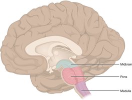
The brain stem consists of the midbrain, pons, and medulla oblongata. It controls vital functions such as heart rate, breathing, and connects the brain to the spinal cord.
Midbrain: Processes visual and auditory information.
Pons: Connects the cerebellum to the brain stem and regulates breathing.
Medulla Oblongata: Controls autonomic functions like heart rate and respiration.
Cerebellum
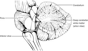
The cerebellum, located posterior to the brain stem, coordinates voluntary movements, balance, and posture. It receives input from the cerebrum and sensory systems to fine-tune motor activity.
Arbor Vitae: The tree-like arrangement of white matter within the cerebellum.
Cranial Nerves
Overview of Cranial Nerves
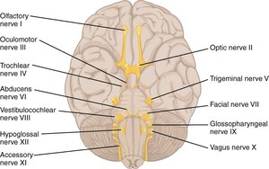
There are twelve pairs of cranial nerves, each with specific sensory, motor, or mixed functions. They primarily serve the head and neck regions, with the vagus nerve extending into the thoracic and abdominal cavities.
Sensory Nerves: Olfactory (I), Optic (II), Vestibulocochlear (VIII)
Motor Nerves: Oculomotor (III), Trochlear (IV), Abducens (VI), Accessory (XI), Hypoglossal (XII)
Mixed Nerves: Trigeminal (V), Facial (VII), Glossopharyngeal (IX), Vagus (X)
Number | Name | Type | Function(s) |
|---|---|---|---|
I | Olfactory | Sensory | Sensory information from the nose (smell) |
II | Optic | Sensory | Sensory information from the eyes (vision) |
III | Oculomotor | Motor | Motor information to most eye muscles |
IV | Trochlear | Motor | Motor information to superior oblique muscle (eye movement) |
V | Trigeminal | Both | Sensory information from and motor information to the face; chewing |
VI | Abducens | Motor | Motor information to lateral rectus muscle (eye movement) |
VII | Facial | Both | Sensory information from anterior tongue; motor to facial muscles; glands |
VIII | Vestibulocochlear | Sensory | Sensory information from the ear (hearing and equilibrium) |
IX | Glossopharyngeal | Both | Sensory from posterior tongue; motor to tongue and throat |
X | Vagus | Both | Sensory from thorax/abdomen; motor to heart, digestive organs, etc. |
XI | Accessory | Motor | Motor information for swallowing and neck muscles |
XII | Hypoglossal | Motor | Motor information to the tongue |
Brain Dissection and Identification
Sheep Brain Dissection
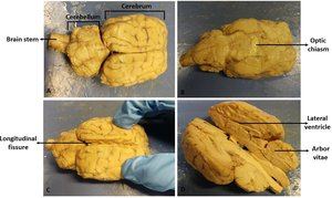
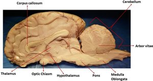
Dissecting a sheep brain allows for hands-on identification of major brain structures, including the cerebrum, cerebellum, brain stem, and internal features such as the corpus callosum and arbor vitae.
Locate external features: cerebrum, cerebellum, longitudinal fissure, gyri, sulci, and lobes.
Identify internal structures: thalamus, hypothalamus, pineal body, pons, medulla, corpus callosum, lateral ventricles, and arbor vitae.
Summary Table: Key Terms and Definitions
Term | Definition | Example/Location |
|---|---|---|
Gray Matter | Regions with many neuron cell bodies and dendrites | Cerebral cortex, basal nuclei |
White Matter | Regions with many myelinated axons | Corpus callosum, internal capsule |
Gyrus | Ridge on the cerebral cortex | Precentral gyrus |
Sulcus | Shallow groove between gyri | Central sulcus |
Fissure | Deep groove in the brain | Longitudinal fissure |
Dura Mater | Outermost, tough meningeal layer | Superficial meningeal layer |
Arachnoid Mater | Middle, web-like meningeal layer | Subarachnoid space contains CSF |
Pia Mater | Innermost, delicate meningeal layer | Adheres to brain surface |
Additional info: The insular lobe, located deep within the lateral sulcus, is involved in motor control, homeostasis, and self-awareness. The cerebellum's arbor vitae is essential for coordinating movement and balance. The cranial nerves are best learned through repeated practice and mnemonic devices.
