 Back
BackCentral Nervous System: Structure, Function, and Integration in Human Anatomy & Physiology
Study Guide - Smart Notes
 Tailored notes based on your materials, expanded with key definitions, examples, and context.
Overview of the Central Nervous System
Introduction to the CNS
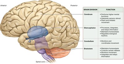
The central nervous system (CNS) is composed of the brain and spinal cord, serving as the primary control center for the body. It integrates sensory information, coordinates voluntary and involuntary responses, and maintains homeostasis.
Brain: Responsible for higher mental functions, sensory interpretation, and initiation of movement.
Spinal Cord: Conducts signals to and from the brain and controls reflex activities.
Basic Structure of the Brain and Spinal Cord
Directional Terms and Landmarks
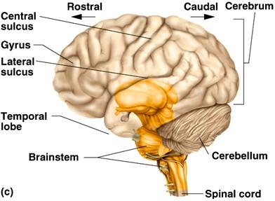
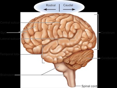
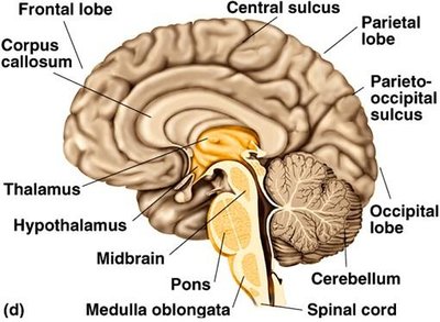
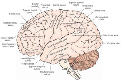
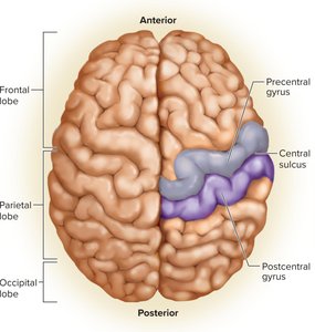
Understanding the orientation and landmarks of the brain is essential for anatomical study. Key terms include rostral (toward the nose/front), caudal (toward the tail/back), gyrus (ridge), and sulcus (groove).
Major Lobes and Surface Anatomy
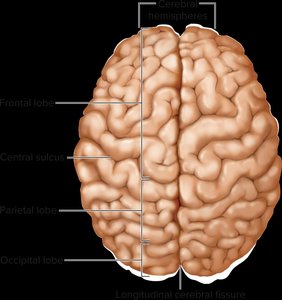
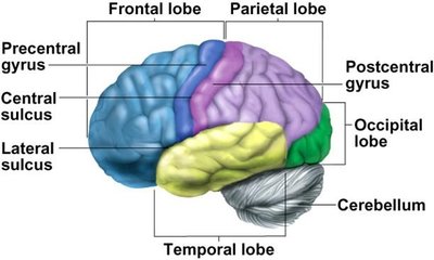
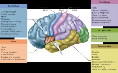
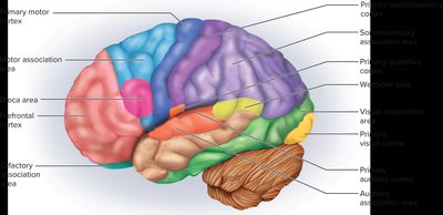
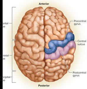
The cerebrum is divided into lobes, each associated with specific functions:
Frontal lobe: Voluntary movement, planning, reasoning
Parietal lobe: Sensory integration
Temporal lobe: Hearing, memory
Occipital lobe: Vision
Internal Brain Anatomy
The brain contains several internal structures, including the thalamus, hypothalamus, midbrain, pons, medulla oblongata, and cerebellum. These regions are responsible for sensory relay, autonomic regulation, and coordination of movement.
CNS White and Gray Matter
Organization of White and Gray Matter
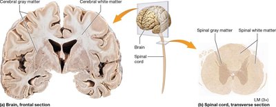
White and gray matter are organized differently in the brain and spinal cord:
Brain: Gray matter forms the outer cortex and inner nuclei; white matter is deeper.
Spinal Cord: White matter is outer; gray matter is deeper.
Gray matter: Contains neuron cell bodies, dendrites, and synapses (unmyelinated).
White matter: Composed of myelinated axons for rapid signal transmission.
Embryonic Development of the CNS
Neurulation and Neural Tube Formation
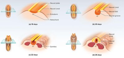
During the first three weeks of embryonic development, the neural plate forms, folds, and fuses to create the neural tube, which gives rise to the CNS. The neural crest forms peripheral nervous system structures.
Neural tube: Becomes the brain and spinal cord.
Neural crest: Forms peripheral nerves and other tissues.
Developmental Regions of the Brain
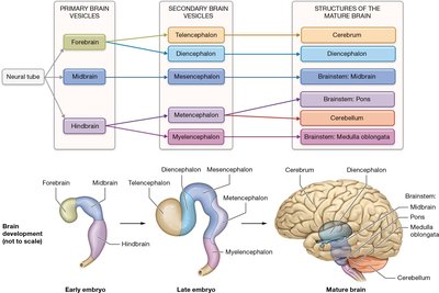
The brain develops from three primary vesicles (forebrain, midbrain, hindbrain), which further differentiate into five secondary vesicles, forming the major adult brain regions.
Primary Vesicle | Secondary Vesicle | Adult Structure |
|---|---|---|
Forebrain | Telencephalon | Cerebrum |
Forebrain | Diencephalon | Diencephalon (thalamus, hypothalamus, epithalamus) |
Midbrain | Mesencephalon | Midbrain |
Hindbrain | Metencephalon | Pons, Cerebellum |
Hindbrain | Myelencephalon | Medulla oblongata |
Cerebrum: Gross Anatomy and Functional Areas
Lobes and Functional Regions
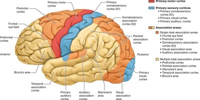
The cerebrum is divided into lobes, each with specialized functions. The precentral gyrus is the primary motor cortex, and the postcentral gyrus is the primary somatosensory cortex.

Cerebral Cortex: Primary and Association Areas
The cerebral cortex contains primary sensory and motor areas, as well as association areas for integration and interpretation of information.
Special and General Senses
Special senses (vision, hearing, taste, smell, equilibrium) and general senses (touch, pressure, pain, temperature) are processed in distinct cortical regions.
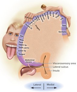
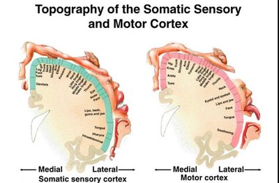
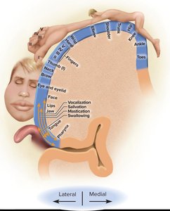
Somatotopic Organization
The primary somatosensory and motor cortices are organized somatotopically, meaning specific regions correspond to specific body parts. This is often illustrated by the sensory and motor homunculus.
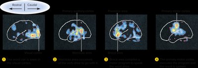
Functional Brain Imaging
Techniques such as PET and fMRI allow visualization of brain activity by detecting changes in blood flow and metabolism.
Basal Nuclei and Cerebral White Matter
Basal Nuclei
The basal nuclei are deep masses of gray matter involved in motor control and inhibition of involuntary movements. They receive input from the substantia nigra and motor cortex and send signals back to these regions.
Cerebral White Matter Tracts
White matter tracts connect different regions of the brain:
Projection tracts: Connect cortex with lower brain/spinal cord
Commissural tracts: Connect hemispheres (e.g., corpus callosum)
Association tracts: Connect regions within the same hemisphere
Limbic System
Structure and Function
The limbic system is a loop of cortical structures involved in emotion, motivation, and memory. Major components include the amygdala, hippocampus, fornix, and cingulate gyrus.
Amygdala: Emotion processing
Hippocampus: Memory formation
Diencephalon
Thalamus
The thalamus is the largest part of the diencephalon, acting as a relay station for sensory information and playing a role in motor control and memory.
Hypothalamus
The hypothalamus regulates autonomic functions, hormone secretion, thermoregulation, hunger, thirst, sleep-wake cycles, and emotional responses.
Epithalamus and Subthalamus
The epithalamus includes the pineal gland, which secretes melatonin to regulate sleep-wake cycles. The subthalamus is involved in motor control and is functionally connected to the basal nuclei.
Cerebellum
Structure and Function
The cerebellum coordinates voluntary movements, balance, posture, and fine motor control. It also contributes to cognitive and emotional processing.
Folia: Parallel folds of gray matter
Vermis: Central region
Lateral hemispheres: Main cerebellar regions
Cerebellar Peduncles
Three pairs of cerebellar peduncles connect the cerebellum to the brainstem, allowing communication with other brain regions.
Brainstem
Major Divisions and Functions
The brainstem connects the spinal cord to the brain and consists of the medulla oblongata, pons, and midbrain. It regulates vital functions such as heart rate, respiration, and reflexes.
Medulla oblongata: Controls autonomic functions and reflexes
Pons: Relays signals, regulates sleep and respiration
Midbrain: Processes visual and auditory information, controls movement
Homeostasis: Role of the Brain
Nervous vs. Endocrine System
Both the nervous and endocrine systems maintain homeostasis, but differ in speed, duration, and specificity of response.
Feature | Nervous System | Endocrine System |
|---|---|---|
Mode of Transport | Axons | Blood |
Speed | Milliseconds | Seconds to days |
Duration | Short | Long |
Area of Effect | Specific | Widespread |
Examples of Homeostatic Regulation
Autonomic nervous system: Hypothalamus regulates heart rate, blood pressure, digestion, and urination.
Body temperature and feeding: Hypothalamus acts as a thermostat and regulates hunger/satiety.
Sleep and wakefulness: Controlled by interactions between cortex, thalamus, hypothalamus, and reticular formation.
Higher Mental Functions
Cognition and Learning
Cognition includes sensory perception, thought, reasoning, judgment, memory, and imagination. Different cortical areas are responsible for these functions, and damage can lead to specific deficits.
Neuroplasticity
Neuroplasticity is the brain's ability to reorganize and form new neural connections. It is enhanced by engaging both hemispheres, practicing new skills, exposure to novelty, meditation, and exercise.
Lateralization of Cerebral Functions
The right and left hemispheres of the brain have specialized functions. The left hemisphere is typically dominant for language and mathematics, while the right is involved in spatial and creative tasks.
Language Centers
Language involves Broca's area (motor speech) and Wernicke's area (language comprehension). Damage to these areas results in different types of aphasia.
Memory
Sensory memory: Lasts less than a second
Short-term memory: Seconds to minutes
Long-term memory: Hours to lifetime; includes declarative (facts) and procedural (skills) memory
Protection of the Brain
Meninges and Spaces
The brain is protected by three meninges: dura mater, arachnoid mater, and pia mater. Spaces between these layers contain blood vessels, CSF, and fat.
Cerebrospinal Fluid (CSF)
CSF is produced by the choroid plexuses, circulates through the ventricles, and is reabsorbed into the bloodstream. It provides buoyancy, protection, and chemical stability for the CNS.
Blood-Brain Barrier
The blood-brain barrier regulates the passage of substances from the blood into the brain, protecting neurons from toxins and fluctuations in blood composition.
The Spinal Cord
Gross Anatomy and Cross-Section
The spinal cord is segmented and gives rise to 31 pairs of spinal nerves. It contains white matter (ascending and descending tracts) and gray matter (neuronal cell bodies).
Ascending and Descending Tracts
Ascending tracts: Sensory pathways to the brain (e.g., spinothalamic, posterior columns)
Descending tracts: Motor pathways from the brain (e.g., corticospinal, reticulospinal)
Motor Neuron Diseases
Poliomyelitis: Destroys motor neurons, leading to paralysis
Multiple sclerosis: Immune attack on myelin sheath
Amyotrophic lateral sclerosis (ALS): Degeneration of motor neurons
Sensation and Movement
Sensory Receptors and Pathways
Sensory receptors detect modality, location, intensity, and duration of stimuli. Sensory pathways involve first-, second-, and third-order neurons relaying information to the cortex.
Pain and Modulation
Pain can be somatic, visceral, or neuropathic. The CNS can modulate pain signals through endogenous opioids and spinal gating mechanisms.
Voluntary Movement
Voluntary movement is coordinated by the cerebral cortex, basal nuclei, and cerebellum. Upper and lower motor neurons transmit signals from the brain to muscles. Decussation (crossing) of pathways explains the effects of brain and spinal cord injuries.
