Back
BackChapter 1: Foundations – Introduction to Human Anatomy and Terminology
Study Guide - Smart Notes
 Tailored notes based on your materials, expanded with key definitions, examples, and context.
Introduction to Human Anatomy
Overview of Human Anatomy
The study of human anatomy involves the examination of both external and internal structures of the human body, as well as the physical relationships between these structures. Anatomy can be studied at various levels, from microscopic (histology and cytology) to macroscopic (gross anatomy), each providing unique insights into the organization and function of the body.
Microscopic Anatomy: Study of structures too small to be seen with the naked eye, such as cells and tissues.
Macroscopic (Gross) Anatomy: Study of structures visible to the naked eye, such as organs and organ systems.
Levels of Biological Organization
Hierarchy of Structural Organization
The human body is organized into a hierarchy of structural levels, each building upon the previous one. Understanding these levels is fundamental to the study of anatomy and physiology.
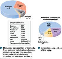
Molecular Level: Involves atoms and molecules essential for life. Four major elements make up over 99% of the human body: hydrogen, oxygen, nitrogen, and carbon.
Major Classes of Molecules: Water, proteins, lipids, and carbohydrates are the primary molecular components.
Cellular Level: The cell is the smallest living unit in the body.
Tissue Level: Four basic tissue types: epithelial, connective, muscle, and nervous tissue.
Organ Level: Structures composed of two or more tissue types working together.
Organ System Level: Groups of organs that perform related functions.
Organism Level: The complete living human being.
Organ System Level
Overview of Major Organ Systems
Organ systems work together to maintain homeostasis and overall health. Each system has specialized functions but interacts closely with others.
Integumentary System: Provides protection and temperature regulation. Includes skin, hair, sweat glands, nails, and sensory receptors.
Skeletal System: Supports the body, stores minerals, and forms blood cells. Divided into axial (skull, vertebral column) and appendicular (limbs, pelvis, shoulder) components.
Muscular System: Responsible for movement, support, and heat production. Includes axial (posture) and appendicular (movement) muscles.
Nervous System: Enables rapid, short-term communication. Central nervous system (CNS: brain and spinal cord) integrates information; peripheral nervous system (PNS) links CNS to other systems.
Endocrine System: Provides long-term communication via hormones. Includes glands such as pineal, pituitary, thyroid, parathyroid, and reproductive organs.
Cardiovascular System: Transports cells and dissolved materials. Composed of the heart, blood, and blood vessels.
Lymphatic System: Manages fluid balance and immune responses. Includes lymph, lymph nodes, thymus, and spleen.
Respiratory System: Facilitates gas exchange. Includes lungs, trachea, larynx, and alveoli.
Digestive System: Breaks down food and absorbs nutrients. Includes stomach, esophagus, intestines, liver, pancreas, salivary glands, and pharynx.
Urinary System: Eliminates waste, regulates salt and water balance, and maintains pH. Includes kidneys, ureters, urinary bladder, and urethra.
Reproductive System: Produces sex cells and hormones. Male: testes; Female: ovaries.
Terminology in Anatomy
Anatomical Position and Regions
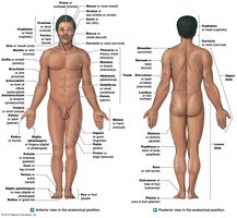
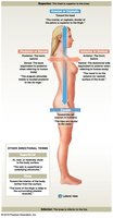
Standardized anatomical terminology is essential for clear communication. The anatomical position is the reference posture for describing locations and directions on the body.
Anatomical Position: Body standing upright, facing forward, arms at sides with palms facing forward.
Supine: Lying face up.
Prone: Lying face down.
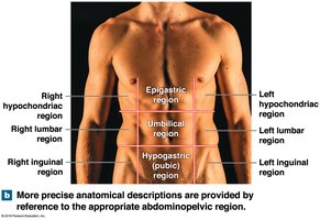
Anatomical Regions and Quadrants
The body is divided into regions and quadrants to facilitate precise anatomical descriptions, especially in clinical settings.
Abdominopelvic Quadrants: Four quadrants (right upper, left upper, right lower, left lower) used to localize pain or pathology.
Abdominopelvic Regions: Nine regions (e.g., epigastric, umbilical, hypogastric) provide more detailed localization.
Anatomical Directions
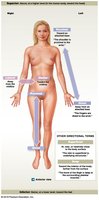
Directional terms describe the locations of structures relative to other structures or locations in the body.
Superior (cranial): Toward the head.
Inferior (caudal): Toward the feet.
Dorsal (posterior): Toward the back.
Ventral (anterior): Toward the front.
Medial: Toward the midline.
Lateral: Away from the midline.
Proximal: Closer to the point of attachment.
Distal: Farther from the point of attachment.
Sectional Anatomy: Body Planes
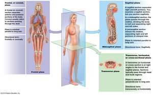
Sectional anatomy refers to the division of the body into planes for study and imaging. The three primary planes are:
Sagittal Plane: Divides the body into right and left portions. Midsagittal is exactly at the midline; parasagittal is offset from the midline.
Frontal (Coronal) Plane: Divides the body into anterior (front) and posterior (back) portions.
Transverse (Horizontal) Plane: Divides the body into superior (upper) and inferior (lower) portions.
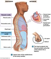
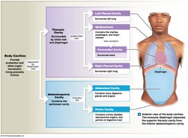
Body Cavities
Major Body Cavities and Their Functions
Body cavities protect internal organs, absorb shock, and allow organs to change shape and size. The two main cavities are dorsal and ventral, each with subdivisions.
Dorsal Cavity: Contains the cranial cavity (brain) and spinal cavity (spinal cord).
Ventral Cavity: Divided by the diaphragm into thoracic and abdominopelvic cavities.
Thoracic Cavity: Contains pleural cavities (lungs), mediastinum, and pericardial cavity (heart).
Abdominopelvic Cavity: Contains abdominal (digestive organs) and pelvic (urinary and reproductive organs) cavities.
Membranes of the Ventral Cavity
Serous Membranes and Their Organization
Serous membranes line the ventral body cavities and cover the organs within them, reducing friction and providing protection. Each cavity has specific membranes:
Pleurae (Lungs): Parietal pleura lines the cavity; visceral pleura covers the lungs.
Pericardium (Heart): Parietal pericardium lines the cavity; visceral pericardium (epicardium) covers the heart.
Peritoneum (Abdominal Organs): Parietal peritoneum lines the cavity; visceral peritoneum covers the organs. Mesenteries are double sheets that support some organs.