 Back
BackChapter 1: The Human Body—An Orientation (Anatomy & Physiology Study Notes)
Study Guide - Smart Notes
 Tailored notes based on your materials, expanded with key definitions, examples, and context.
Chapter 1: The Human Body—An Orientation
Introduction to Anatomy and Physiology
Anatomy and physiology are foundational sciences for understanding the structure and function of the human body. Anatomy focuses on the form and relationships of body parts, while physiology explores how these parts work to sustain life.
Anatomy: Study of the structure of body parts and their relationships to one another.
Physiology: Study of the function of body parts; how they carry out life-sustaining activities.
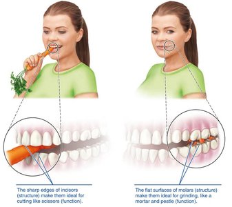
Principle of Complementarity: Structure determines function; what a structure can do depends on its specific form.
Reference Standards and Anatomical Variability
Textbook values are based on reference standards:
Reference male: Healthy, young male weighing about 70 kg (155 lb).
Reference female: Healthy, young female weighing about 57 kg (125 lb).
Anatomical variability: Over 90% of anatomical structures match textbook descriptions, but minor variations exist (e.g., nerves or blood vessels may be out of place, small muscles may be missing).
Sex vs. Gender
Sex: Biological attributes based on chromosomes, gene expression, and hormone action; reflected in reproductive anatomy and physiology (male or female).
Gender: Psychosocial construct including behaviors, expressions, and identities (e.g., man, woman, transgender, non-binary).
Topics and Subdivisions of Anatomy and Physiology
Subdivisions of Anatomy
Gross (macroscopic) anatomy: Study of large body structures visible to the naked eye.
Regional anatomy: All structures in a particular area.
System anatomy: One system (e.g., cardiovascular, nervous).
Surface anatomy: Internal structures as related to overlying skin.
Microscopic anatomy: Structures too small to be seen with the naked eye.
Cytology: Study of cells.
Histology: Study of tissues.
Developmental anatomy: Structural changes throughout the lifespan.
Embryology: Developmental changes before birth.
Studying Anatomy
Requires understanding anatomical terminology and using observation, manipulation, palpation (feeling organs), and auscultation (listening with a stethoscope).
Medical imaging (e.g., X-ray, MRI, CT, ultrasound) allows non-invasive internal visualization.
Subdivisions of Physiology
Based on organ systems (e.g., renal, neurophysiology, cardiovascular physiology).
Often focuses on cellular or molecular events and chemical/physical principles (e.g., electrical currents, pressure, lever systems).
Levels of Structural Organization in the Human Body
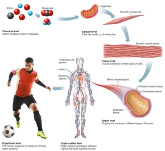
The human body is organized into hierarchical levels, from the simplest chemical components to the entire organism.
Chemical level: Atoms combine to form molecules.
Cellular level: Cells are made up of molecules and organelles.
Tissue level: Tissues consist of similar types of cells.
Organ level: Organs are made up of different types of tissues.
Organ system level: Organ systems consist of different organs that work together closely.
Organismal level: The human organism is made up of many organ systems.
The Body’s Organ Systems and Their Major Functions
The human body contains 11 organ systems, each with specific functions essential for survival.
System | Main Functions |
|---|---|
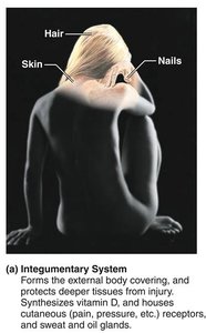
Integumentary | Protects body, synthesizes vitamin D, houses receptors and glands |
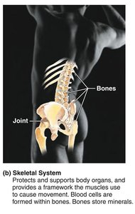
Skeletal | Supports and protects organs, forms blood cells, stores minerals |
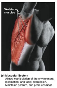
Muscular | Movement, posture, heat production |
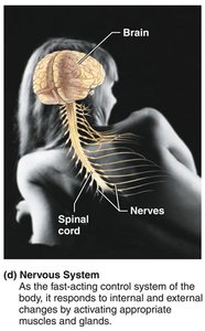
Nervous | Fast-acting control, responds to stimuli |
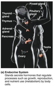
Endocrine | Hormone secretion, regulates growth, metabolism, reproduction |
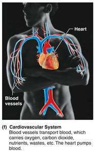
Cardiovascular | Transports blood, nutrients, wastes, gases |
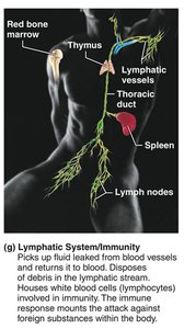
Lymphatic/Immunity | Returns fluid to blood, immune response |
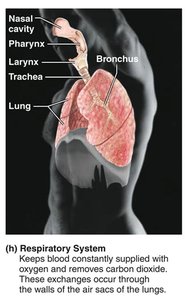
Respiratory | Gas exchange (O2/CO2) |
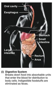
Digestive | Breaks down food, absorbs nutrients, eliminates waste |
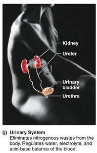
Urinary | Eliminates nitrogenous wastes, regulates water/electrolytes |
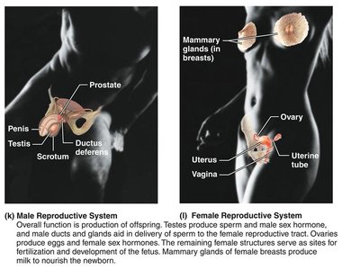
Reproductive | Produces offspring |
Necessary Life Functions
All living organisms must perform certain vital functions to maintain life:
Maintaining boundaries: Separation between internal and external environments (e.g., plasma membranes, skin).
Movement: Of body parts (skeletal muscles), substances (cardiac/smooth muscle), and cells.
Responsiveness: Ability to sense and respond to stimuli (e.g., withdrawal reflex, breathing rate).
Digestion: Breakdown of food and absorption of nutrients.
Metabolism: All chemical reactions in body cells (catabolism, anabolism, cellular respiration).
Excretion: Removal of wastes (e.g., urea, CO2, feces).
Reproduction: Cellular division for growth/repair; organismal production of offspring.
Growth: Increase in size of a body part or organism.
Survival Needs
Humans require several factors for survival, each in appropriate amounts:
Nutrients: Chemicals for energy and cell building (carbohydrates, proteins, fats, vitamins, minerals).
Oxygen: Essential for metabolic reactions; body can survive only a few minutes without it.
Water: Most abundant chemical in the body; necessary for chemical reactions and as a fluid base.
Normal body temperature: Needed for proper metabolic reaction rates (about 37°C or 98.6°F).
Appropriate atmospheric pressure: Required for adequate breathing and gas exchange in the lungs.
Homeostasis and Feedback Mechanisms
Definition and Importance
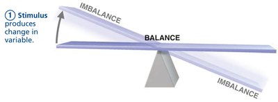
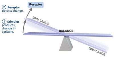
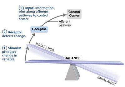
Homeostasis is the maintenance of relatively stable internal conditions despite continuous environmental changes. It is a dynamic equilibrium, maintained by all organ systems, and is essential for health.
Law of mass balance: The amount of a substance taken in must equal the amount lost to maintain constancy.
Homeostatic Control Mechanisms
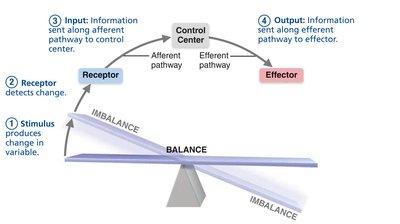
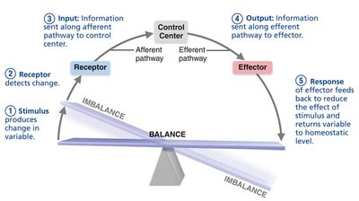
Homeostatic regulation involves three main components:
Receptor (sensor): Monitors the environment and responds to stimuli.
Control center: Determines the set point and appropriate response.
Effector: Provides the means to respond, reducing or enhancing the stimulus.
Negative Feedback
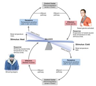
Most common feedback mechanism in the body.
Response reduces or shuts off the original stimulus, returning the variable to its set point.
Examples: Regulation of body temperature, regulation of blood glucose by insulin.
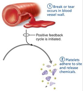
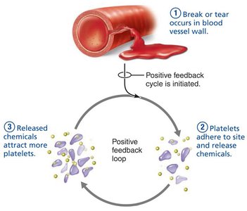
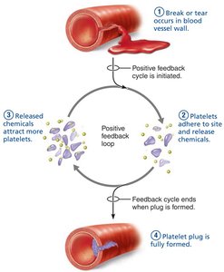
Positive Feedback
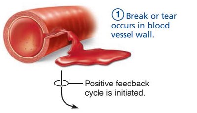
Response enhances or exaggerates the original stimulus.
Usually controls infrequent events that do not require continuous adjustment.
Examples: Enhancement of labor contractions by oxytocin, platelet plug formation and blood clotting.
Feedforward (Anticipatory) Response
Occurs in anticipation of a change to the internal environment (e.g., salivation before eating).
Homeostatic Imbalance
Disturbance of homeostasis increases risk of disease and contributes to aging.
If negative feedback is overwhelmed, destructive positive feedback may occur (e.g., heart failure).
Anatomical Terms: Body Directions, Regions, and Planes
Anatomical Position and Directional Terms
Anatomical position: Body erect, feet slightly apart, palms facing forward, thumbs away from body.
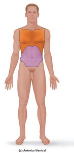
Directional terms describe the location of one body part relative to another (e.g., superior, inferior, anterior, posterior, medial, lateral, proximal, distal, superficial, deep).
Orientation and Directional Terms Table
Term | Definition | Example |
|---|---|---|
Superior (cranial) | Toward the head end or upper part; above | The head is superior to the abdomen. |
Inferior (caudal) | Away from the head end or toward the lower part; below | The navel is inferior to the chin. |
Anterior (ventral) | Toward or at the front; in front of | The breastbone is anterior to the spine. |
Posterior (dorsal) | Toward or at the back; behind | The heart is posterior to the breastbone. |
Medial | Toward or at the midline; on the inner side | The heart is medial to the arm. |
Lateral | Away from the midline; on the outer side | The arms are lateral to the chest. |
Intermediate | Between a more medial and a more lateral structure | The collarbone is intermediate between the breastbone and shoulder. |
Proximal | Closer to the origin of the body part or point of attachment | The elbow is proximal to the wrist. |
Distal | Farther from the origin or point of attachment | The knee is distal to the thigh. |
Superficial (external) | Toward or at the body surface | The skin is superficial to skeletal muscles. |
Deep (internal) | Away from the body surface; more internal | The lungs are deep to the skin. |
Regional Terms
Axial part: Main axis of the body (head, neck, trunk).
Appendicular part: Limbs (arms and legs).
Regional terms designate specific areas within these divisions.
Body Planes and Sections
Sagittal plane: Divides body into right and left parts.
Midsagittal (median): Lies exactly in the midline.
Parasagittal: Offset from the midline.
Frontal (coronal) plane: Divides body into anterior and posterior parts.
Transverse (horizontal) plane: Divides body into superior and inferior parts.
Oblique section: Cuts made at angles other than 90° to the vertical plane.
Body Cavities and Membranes
Major Body Cavities
Dorsal body cavity: Protects the nervous system; includes cranial cavity (brain) and vertebral cavity (spinal cord), covered by meninges.
Ventral body cavity: Houses internal organs (viscera); includes thoracic cavity (pleural cavities, mediastinum, pericardial cavity) and abdominopelvic cavity (abdominal and pelvic cavities).
Membranes in the Ventral Body Cavity
Serosa (serous membrane): Thin, double-layered membrane covering ventral cavity surfaces.
Parietal serosa: Lines cavity walls.
Visceral serosa: Covers organs.
Serous fluid between layers reduces friction.
Examples:
Pleura: Lungs
Pericardium: Heart
Peritoneum: Abdominopelvic organs
Abdominopelvic Regions and Quadrants
Medical personnel use four quadrants: RUQ, LUQ, RLQ, LLQ.
Anatomists use nine regions: umbilical, epigastric, pubic (hypogastric), right/left inguinal (iliac), right/left lateral (lumbar), right/left hypochondriac.
Other Body Cavities
Open to environment: Oral/digestive, nasal, orbital, middle ear cavities.
Closed: Synovial cavities (joints).
Clinical Connections
Hiatal hernia: Stomach protrudes through diaphragm into thoracic cavity, causing heartburn.
Pleurisy/Peritonitis: Inflammation of serous membranes, causing pain and organ dysfunction.
Wrong site surgery: Critical errors due to misunderstanding anatomical terminology (e.g., amputation of wrong limb).
Additional info: Understanding anatomical terminology and body organization is essential for accurate communication in healthcare and for the study of all subsequent chapters in Anatomy & Physiology.
