 Back
BackChapter 11C: Synapses and Neural Integration – Nervous System and Nervous Tissue
Study Guide - Smart Notes
 Tailored notes based on your materials, expanded with key definitions, examples, and context.
IV. The Synapse
Definition and Functional Role
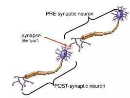
The synapse is a specialized junction that mediates information transfer from one neuron to another, or from a neuron to an effector cell. Synapses are essential for neural communication and integration within the nervous system.
Presynaptic neuron: Conducts impulses toward the synapse and sends information.
Postsynaptic neuron: Transmits electrical signals away from the synapse and receives information.
Types of Synapses
Structural Classification
Synapses can be classified based on their location and the structures they connect:
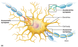
Axodendritic synapses: Between axon terminals of one neuron and dendrites of another.
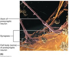
Axosomatic synapses: Between axon terminals and the cell body (soma) of another neuron.
Axoaxonal synapses: Between axon terminals of two neurons.
Chemical Synapses
Structure and Function
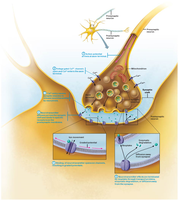
Chemical synapses are the most common type in the nervous system. They use neurotransmitters to transmit signals across a fluid-filled synaptic cleft.
Axon terminal: Contains synaptic vesicles filled with neurotransmitter.
Receptor region: Located on the postsynaptic neuron's membrane, usually on dendrites or cell body.
Synaptic cleft: The gap separating the presynaptic and postsynaptic membranes.
Steps of Chemical Synaptic Transmission
The process of neurotransmitter release and signal transmission involves several steps:
Action potential arrives at the axon terminal.
Voltage-gated Ca2+ channels open, allowing Ca2+ influx.
Ca2+ triggers synaptic vesicles to release neurotransmitter by exocytosis.
Neurotransmitter diffuses across the synaptic cleft and binds to receptors on the postsynaptic membrane.
Binding opens ion channels, resulting in graded potentials.
Neurotransmitter effects are terminated by reuptake, enzymatic degradation, or diffusion away from the synapse.
Neurotransmitters
Types and Effects
Neurons can produce two or more neurotransmitters, allowing them to exert multiple influences. The effect of a neurotransmitter depends on the type of receptor it binds to:
Excitatory (depolarizing): Promotes action potential generation.
Inhibitory (hyperpolarizing): Suppresses action potential generation.
Example: Acetylcholine (ACh) is excitatory at neuromuscular junctions in skeletal muscle, but inhibitory in cardiac muscle.
Summation
Integration of Synaptic Inputs
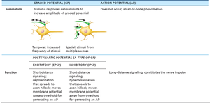
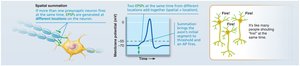
A single excitatory postsynaptic potential (EPSP) cannot induce an action potential (AP). EPSPs and inhibitory postsynaptic potentials (IPSPs) can summate to influence whether an AP is generated. Most neurons receive both excitatory and inhibitory inputs from thousands of other neurons.
Temporal summation: One or more presynaptic neurons transmit impulses in rapid-fire order.
Spatial summation: Multiple presynaptic neurons transmit impulses simultaneously at different locations.
Threshold: Only if EPSPs predominate and bring the membrane potential to threshold will an AP be generated.
Receptors
Channel-Linked Receptors
Channel-linked receptors mediate rapid synaptic transmission by directly opening ion channels upon neurotransmitter binding.
Fast response: Allows quick changes in membrane potential.
Example: Ligand-gated ion channels for Na+ or K+.
G Protein–Linked Receptors
G protein–linked receptors cause the formation of intracellular second messengers, leading to slower but longer-lasting effects.
Indirect response: Activates signaling cascades inside the cell.
Example: cAMP pathway.
VI. Neural Integration
Neuronal Pools and Integration
Neurons function together in groups called neuronal pools, which contribute to broader neural functions. Integration ensures that billions of neurons in the CNS operate smoothly as a whole.
Neuronal pool: Functional group of neurons that process and relay information.
A Simple Reflex Arc
A reflex arc is a basic neural circuit that mediates rapid, automatic responses to stimuli. It typically involves sensory input, integration in the CNS, and motor output.
Components: Receptor, sensory neuron, integration center, motor neuron, effector.
Types of Circuits in Neuronal Pools
Neuronal pools can be organized into different types of circuits, each with distinct functional properties:
Diverging circuit: One input, many outputs; amplifies signal.
Converging circuit: Many inputs, one output; concentrates signal.
Reverberating circuit: Chain of neurons with feedback; produces rhythmic activity.
Parallel after-discharge circuit: Several pathways; produces bursts of activity.
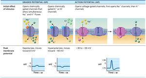
Summary Table: Graded Potential vs. Action Potential
This table compares the properties of graded potentials and action potentials, as well as the functions of EPSPs and IPSPs.
Property | Graded Potential (GP) | Action Potential (AP) |
|---|---|---|
Summation | Multiple responses can summate to increase amplitude | Does not occur; all-or-none phenomenon |
Initial effect of stimulus | Opens chemically gated channels (Na+ and K+ fluxes) | Opens voltage-gated channels (Na+ then K+) |
Peak membrane potential | Depolarizes (moves toward 0 mV) or hyperpolarizes (moves toward -90 mV) | +30 to -50 mV |
Function | Short-distance signaling; depolarization (EPSP) or hyperpolarization (IPSP) | Long-distance signaling; constitutes the nerve impulse |
Additional info: The notes above expand on the original content by providing definitions, examples, and context for synaptic transmission, neurotransmitter effects, summation, and neural integration. The included images directly reinforce the explanations of synapse structure, function, and summation.
