 Back
BackChapter 14: The Autonomic Nervous System – Structure, Function, and Clinical Relevance
Study Guide - Smart Notes
 Tailored notes based on your materials, expanded with key definitions, examples, and context.
The Autonomic Nervous System (ANS)
Overview and Importance
The autonomic nervous system (ANS) is a critical component of the nervous system responsible for regulating involuntary functions such as heart rate, blood pressure, digestion, and glandular activity. Understanding the ANS is essential for anticipating the effects and side effects of drugs on patients.
Structural Organization of the Nervous System
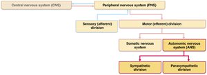
The nervous system is divided into the central nervous system (CNS) and peripheral nervous system (PNS). The PNS is further subdivided into sensory (afferent) and motor (efferent) divisions. The motor division includes the somatic nervous system (controls skeletal muscles) and the autonomic nervous system (controls smooth muscle, cardiac muscle, and glands).
Comparison: Autonomic vs. Somatic Nervous System
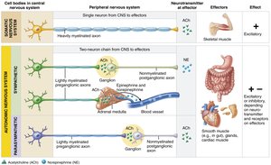
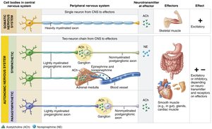
Both systems have motor fibers but differ in effectors, number of neurons between CNS and effectors, and neurotransmitter actions.
Somatic Nervous System: Innervates skeletal muscles; uses a single, thick myelinated axon; neurotransmitter is acetylcholine (ACh), always excitatory.
Autonomic Nervous System: Innervates cardiac muscle, smooth muscle, and glands; uses a two-neuron chain (preganglionic and postganglionic); neurotransmitters include ACh and norepinephrine (NE), effects can be excitatory or inhibitory.
Divisions of the Autonomic Nervous System
Parasympathetic and Sympathetic Divisions
The ANS consists of two main divisions:
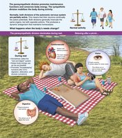
Parasympathetic Division: Promotes maintenance functions and conserves energy; known as the "rest-and-digest" system.
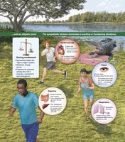
Sympathetic Division: Mobilizes the body during activity; known as the "fight-or-flight" system.
Most visceral organs are served by both divisions, which have opposing effects to maintain homeostasis.
Functional Roles
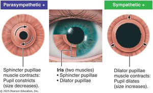
Parasympathetic: Directs digestion, diuresis, and defecation. During relaxation, heart rate and blood pressure are low, gastrointestinal activity is high, and pupils are constricted.
Sympathetic: Activated during exercise, excitement, or emergency. Increases heart rate, dilates pupils and bronchioles, shunts blood to muscles, and causes liver to release glucose.
Anatomical Differences
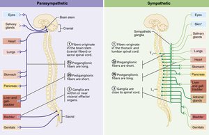
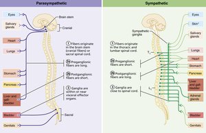
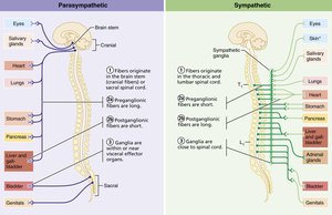
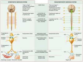
Sites of Origin: Parasympathetic fibers are craniosacral; sympathetic fibers are thoracolumbar.
Fiber Lengths: Parasympathetic has long preganglionic and short postganglionic fibers; sympathetic has short preganglionic and long postganglionic fibers.
Ganglia Location: Parasympathetic ganglia are near or within effectors; sympathetic ganglia are close to the spinal cord.
Parasympathetic Division
Cranial and Sacral Components
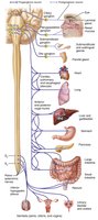
The parasympathetic division is also called the craniosacral division. Preganglionic fibers originate from the brainstem and sacral spinal cord, synapsing in terminal ganglia near or within target organs.
Cranial Nerves and Functions
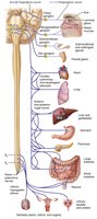
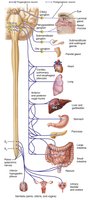
Oculomotor (III): Controls eye muscles, pupil constriction, and lens accommodation.
Facial (VII): Stimulates glands in the head.
Glossopharyngeal (IX): Stimulates parotid salivary glands.
Vagus (X): Accounts for ~90% of parasympathetic fibers; serves thoracic and abdominal viscera.
Sympathetic Division
Thoracolumbar Origin and Complexity
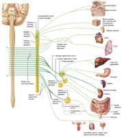
The sympathetic division originates from spinal cord segments T1–L2 and innervates more organs than the parasympathetic division. Some structures, such as sweat glands and blood vessels, are innervated only by the sympathetic division.
Sympathetic Trunks and Ganglia
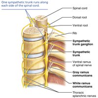
Preganglionic fibers pass through white rami communicantes to enter sympathetic trunk ganglia, which flank each side of the vertebral column. There are typically 23 ganglia per trunk.
Pathways of Sympathetic Innervation
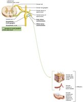
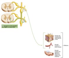
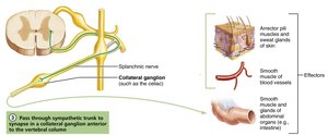
Synapse at the same level: Preganglionic and postganglionic neurons synapse in the same trunk ganglion.
Synapse at a higher or lower level: Preganglionic axon ascends or descends to another trunk ganglion.
Synapse in collateral ganglion: Preganglionic axon passes through trunk ganglion and synapses in collateral ganglia anterior to the vertebral column.
Summary Table: Autonomic Ganglia
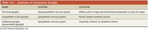
The table below summarizes the main types of autonomic ganglia, their division, and location.
NAME | DIVISION | LOCATION |
|---|---|---|
Terminal ganglia | Parasympathetic nervous system | Within wall of organ served (intramural ganglia) or close to organ |
Sympathetic trunk ganglia | Sympathetic nervous system | Paired, beside vertebral column |
Collateral ganglia (prevertebral ganglia) | Sympathetic nervous system | Unpaired, anterior to vertebral column |
Sympathetic Pathways to Organs
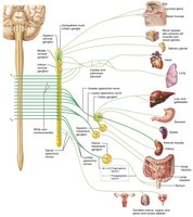
Head: Fibers emerge from T1–T4, synapse in superior cervical ganglion, innervate skin, blood vessels, iris, glands, and heart.
Thorax: Fibers emerge from T1–T6, synapse in cervical trunk ganglia, innervate heart, thyroid, skin, lungs, and esophagus.
Abdomen: Fibers from T5–L2 travel through splanchnic nerves, synapse in celiac and mesenteric ganglia, serve digestive organs.
Pelvis: Fibers from T10–L2 serve pelvic organs, inhibit activity of muscles and glands.
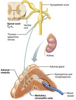
Adrenal Medulla: Preganglionic fibers stimulate chromaffin cells to release epinephrine and norepinephrine.
Visceral Reflex Arcs
Components and Differences
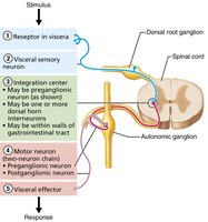
Visceral reflex arcs regulate involuntary functions and differ from somatic reflex arcs in having two consecutive neurons in the motor pathway, visceral sensory afferents, and effectors being smooth muscle, cardiac muscle, or glands.
Neurotransmitters and Receptors
Major Neurotransmitters
Acetylcholine (ACh): Released by all ANS preganglionic axons and all parasympathetic postganglionic axons.
Norepinephrine (NE): Released by most sympathetic postganglionic axons.
The effect depends on the type of receptor (cholinergic or adrenergic) present on the target organ.
Cholinergic Receptors
Nicotinic receptors: Found on all postganglionic neurons, adrenal medulla, and skeletal muscle cells; always excitatory.
Muscarinic receptors: Found on all effector cells stimulated by postganglionic cholinergic fibers; can be excitatory or inhibitory depending on the organ.
Adrenergic Receptors
Alpha (α) receptors: Subclasses α1 and α2.
Beta (β) receptors: Subclasses β1, β2, and β3.
Effects depend on which subclass predominates on the target organ.
Parasympathetic and Sympathetic Interactions
Dual Innervation and Antagonism
Most organs receive input from both divisions, which interact antagonistically to maintain precise control of visceral activity. Sympathetic increases heart rate and inhibits digestion; parasympathetic decreases heart rate and promotes digestion.
Sympathetic and Parasympathetic Tone
Sympathetic tone: Maintains partial constriction of blood vessels; can increase or decrease blood pressure as needed.
Parasympathetic tone: Dominates heart and digestive/urinary organs; slows heart rate and promotes normal activity.
Cooperative Effects
Some functions, such as control of external genitalia, require cooperation between both divisions: parasympathetic causes erection, sympathetic causes ejaculation.
Unique Roles and Effects of the Sympathetic Division
Thermoregulation: Controls sweat glands and blood vessel dilation/constriction.
Renin Release: Stimulates kidneys to release renin, affecting blood pressure.
Metabolic Effects: Increases metabolic rate, blood glucose, and mobilizes fats.
Localized vs. Diffuse Effects
Parasympathetic: Short-lived, localized effects due to rapid breakdown of ACh.
Sympathetic: Longer-lasting, bodywide effects due to slower inactivation of NE and hormone release.
Control of ANS Activity
CNS Centers
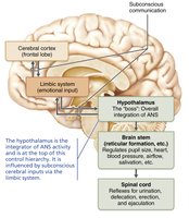
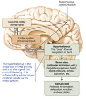
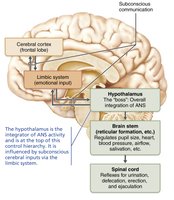
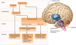
The ANS is regulated by the brainstem, spinal cord, hypothalamus, and cerebral cortex. The hypothalamus is the main integrative center, with input from the limbic system and cerebral cortex.
Disorders of the Autonomic Nervous System
Common Disorders
Hypertension: Overactive sympathetic response; treated with adrenergic blockers.
Autonomic dysreflexia: Uncontrolled activation in spinal cord injury patients; risk of stroke.
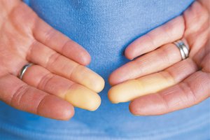
Raynaud’s disease: Exaggerated vasoconstriction in fingers and toes; treated with vasodilators.
Postural Orthostatic Tachycardia Syndrome (POTS)
POTS is characterized by an abnormal increase in heart rate upon standing, accompanied by vasodilation and low blood pressure. Symptoms include dizziness, fatigue, and cold extremities. Treatment involves dietary changes, exercise, and medications to block sympathetic receptors.
Developmental Aspects of the ANS
Origin and Aging
Development: ANS preganglionic neurons derive from the neural tube; postganglionic neurons and ganglia from the neural crest.
Aging: ANS efficiency declines with age, leading to constipation, dry eyes, orthostatic hypotension, and less responsive pressure receptors.
