 Back
BackChapter 20: The Lymphatic System and Immunity – Structured Study Notes
Study Guide - Smart Notes
 Tailored notes based on your materials, expanded with key definitions, examples, and context.
The Lymphatic System and Immunity
Overview of the Lymphatic System
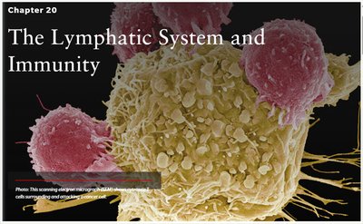
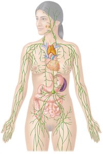
The lymphatic system is a vital component of the body's defense mechanisms, working closely with the immune system to protect against disease and maintain fluid balance. It consists of lymphatic vessels, lymphoid tissues, and organs that drain interstitial fluid, transport dietary fats, and facilitate immune responses.
Lymphatic vessels collect excess interstitial fluid and return it to the bloodstream.
Lymphoid tissues and organs include lymph nodes, spleen, thymus, and mucosa-associated lymphoid tissue (MALT).
Functions: Fluid drainage, fat transport, and immune protection.
Lymphatic Circulation vs. Systemic Circulation
Lymphatic circulation differs from systemic (blood) circulation in several key ways. Both systems return fluid toward the heart, but lymphatic vessels carry lymph, not blood, and lack a central pump.
Similarities: Both return fluid to the heart and use valves to prevent backflow.
Differences: Lymphatic vessels carry lymph, veins carry blood; lymphatic system relies on skeletal muscle and respiratory pumps, not a heart.
Structure and Function of Lymphatic Vessels
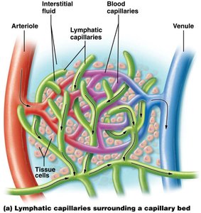
Lymphatic vessels begin as blind-ended capillaries in tissues, collecting excess interstitial fluid. This fluid, now called lymph, is transported through larger vessels and trunks, eventually draining into the subclavian veins.
Lymphatic capillaries are highly permeable and allow entry of fluid, proteins, and immune cells.
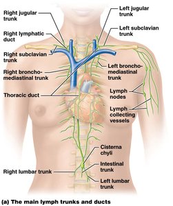
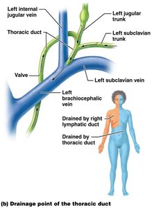
Principal lymphatic trunks: Five main trunks unite into two channels: the right lymphatic duct and thoracic duct.
Drainage: Right lymphatic duct drains the right upper body; thoracic duct drains the rest.
Fluid Dynamics and Edema
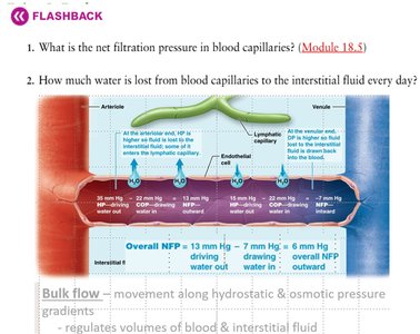
More fluid leaves blood capillaries than returns, which could lead to edema if not drained by the lymphatic system. Net filtration pressure and bulk flow regulate fluid movement.
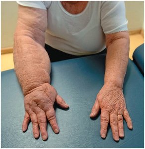
Edema: Occurs when lymphatic drainage is impaired, leading to tissue swelling.
Bulk flow: Driven by hydrostatic and osmotic pressure gradients.
Lymphatic Capillary Structure and Function
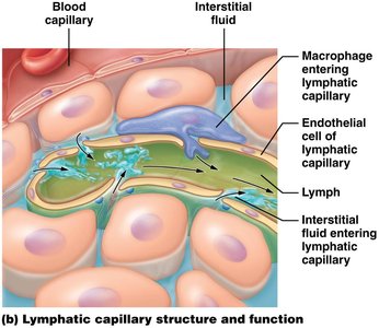
Lymphatic capillaries surround capillary beds and collect interstitial fluid, which becomes lymph. Their unique structure allows for the entry of large molecules and immune cells.
Endothelial cells overlap to form one-way valves.
Macrophages and other immune cells can enter lymphatic capillaries.
Lymphatic Organs and Tissues
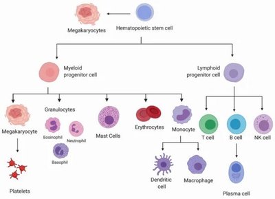
The lymphatic system includes primary and secondary organs. Primary organs (thymus and red bone marrow) are sites of lymphocyte maturation; secondary organs (lymph nodes, spleen, MALT) are sites of immune function.
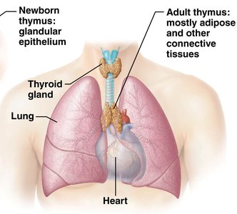
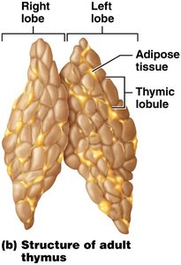
Thymus: Maturation site for T lymphocytes; largest in childhood.
Red bone marrow: Site of B lymphocyte maturation.
Lymph nodes: Filter lymph and destroy foreign substances.
Lymph nodules (MALT): Unencapsulated clusters in mucous membranes (tonsils, Peyer's patches).
Spleen: Largest lymphoid organ; filters blood, destroys old RBCs, and mounts immune responses.
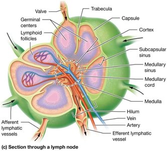
Microscopic Structure of Lymphoid Organs
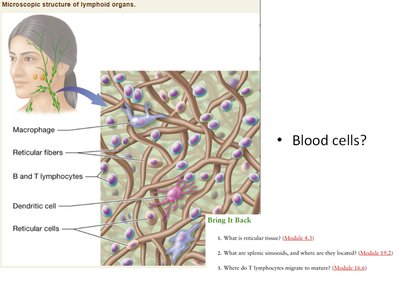
Lymphoid organs contain reticular fibers, macrophages, dendritic cells, and lymphocytes. These structures support immune cell function and filtration.
Reticular tissue: Forms the structural framework.
Macrophages and dendritic cells: Present antigens and initiate immune responses.
Immunity: Innate and Adaptive
Immunity is divided into innate (non-specific) and adaptive (specific) responses. Innate immunity provides immediate, general protection; adaptive immunity targets specific pathogens and develops memory.
Innate immunity: Includes surface barriers, cells, and proteins.
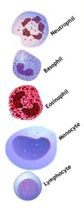
Adaptive immunity: Involves lymphocytes (T and B cells), antibodies, and memory cells.
Innate Immunity: First and Second Lines of Defense
First line: Physical (skin, mucous membranes), chemical (secretions), and mechanical (expulsion) barriers.
Second line: Antimicrobial proteins (interferons, complement), natural killer cells, fever, phagocytosis, and inflammation.
Phagocytosis
Neutrophils and macrophages ingest and digest invaders.
Three phases: chemotaxis, adherence, ingestion.
Inflammation
Four cardinal signs: redness, swelling, pain, heat.
Three stages: vasodilation, phagocyte migration, tissue repair.
Fever
Elevated body temperature inhibits microbial growth and enhances tissue repair.
Adaptive Immunity: Cell-Mediated and Antibody-Mediated Responses
Adaptive immunity is characterized by specificity and memory. It is divided into cell-mediated (T cells) and antibody-mediated (B cells) responses.
Cell-mediated: T lymphocytes destroy infected or abnormal cells.
Antibody-mediated: B lymphocytes produce antibodies to neutralize pathogens.
Clonal selection: Proliferation and differentiation of lymphocytes upon antigen exposure.
Antigens and Antigen Processing
Antigens: Foreign substances recognized by the immune system.
Antigenic determinants (epitopes): Specific regions that provoke immune responses.
MHC proteins: Mark cells as "self" and present antigens to T cells.
Endogenous antigens: Synthesized within the body (e.g., viral proteins).
Exogenous antigens: From outside the body, processed by antigen-presenting cells (APCs).
T Cell Activation
Helper T cells (Th): Recognize antigens from MHC-II, secrete cytokines.
Cytotoxic T cells (Tc): Recognize antigens from MHC-I, destroy infected cells via perforin and granzymes.
B Cell Activation and Antibody Structure
B cells bind antigens, receive costimulation from Th cells, and differentiate into plasma cells (secrete antibodies) and memory cells.
Antibody structure: Y-shaped, four polypeptide chains (two heavy, two light), variable and constant regions.
Classes of Immunoglobulins
Class | Structure | Function |
|---|---|---|
IgG | Monomer | Main antibody in blood; crosses placenta |
IgA | Dimer | Found in secretions; localized protection |
IgM | Pentamer | First antibody produced; blood typing |
IgE | Monomer | Allergies and hypersensitivity |
IgD | Monomer | B cell activation |
Immunological Memory
Memory cells provide a faster, stronger response upon second exposure to the same antigen.
Primary response: First exposure, slower antibody production.
Secondary response: Subsequent exposure, rapid and robust antibody production (especially IgG).
Immune Disorders
Autoimmune diseases: Immune system attacks self-tissues (e.g., Grave's disease, Hashimoto's thyroiditis).
Immunocompromised: Reduced immune function.
Hypersensitivity: Allergic reactions mediated by IgE.
Clinical Application: Lymphedema
Lymphedema is a condition characterized by increased fluid retention and tissue swelling due to lymphatic system pathology.
Causes: Removal of lymph nodes, impaired lymphatic drainage.
Symptoms: Swelling, especially in limbs.
Summary Table: Lymphatic Organs
Organ | Function | Location |
|---|---|---|
Thymus | T cell maturation | Between sternum and heart |
Red bone marrow | B cell maturation | Within bones |
Lymph nodes | Filter lymph | Along lymphatic vessels |
Spleen | Filter blood, destroy old RBCs | Inferior to stomach |
MALT | Immune surveillance | Mucous membranes |
Key Equations
Net Filtration Pressure (NFP):
Additional info: Academic context was added to clarify immune cell functions, lymphatic organ roles, and clinical relevance of lymphedema.
