 Back
BackChapter 22: The Respiratory System – Anatomy & Physiology II Study Notes
Study Guide - Smart Notes
 Tailored notes based on your materials, expanded with key definitions, examples, and context.
Functional Anatomy of the Respiratory System
Overview of the Respiratory System
The respiratory system is responsible for the exchange of gases (oxygen and carbon dioxide) between the atmosphere and the blood. It is divided into upper and lower respiratory tracts, each with specialized structures and functions.
Upper respiratory tract: Nose, nasal cavity, paranasal sinuses, and pharynx
Lower respiratory tract: Larynx, trachea, bronchi, bronchioles, and lungs (including alveoli)
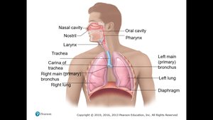
Main Functions of the Respiratory System
Air conduction: Provides a passageway for air to move into and out of the lungs
Air filtration: Filters, warms, and moistens incoming air
Gas exchange: Facilitates the exchange of O2 and CO2 between air and blood
Speech: Houses structures necessary for vocalization
Olfaction: Contains receptors for the sense of smell
Upper Respiratory System
The Nose and Paranasal Sinuses
The nose is the only external part of the respiratory system and serves as the main entrance for air. It is divided into the external nose and the nasal cavity.
Functions of the nose:
Provides an airway for respiration
Moistens and warms entering air
Filters and cleans inspired air
Serves as a resonating chamber for speech
Houses olfactory receptors
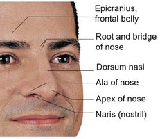
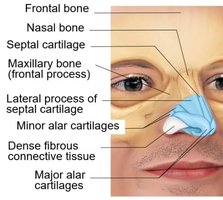
Nasal Cavity
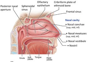
The nasal cavity is lined with respiratory mucosa containing goblet cells and ciliated epithelium. The cilia sweep contaminated mucus toward the throat, preventing particles from reaching the lungs. The conchae increase the surface area and enhance air turbulence, aiding in filtering, warming, and moistening air during inhalation, and reclaiming heat and moisture during exhalation.
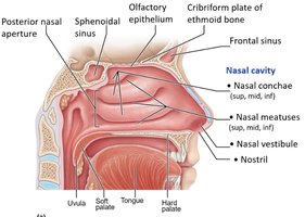
Clinical Note: Rhinitis and Sinusitis
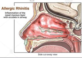
Rhinitis is the inflammation of the nasal mucosa, which can spread to the throat and chest due to the continuity of the mucosa. Blockage of sinus passageways can result in sinusitis, causing headaches due to air absorption.
The Pharynx
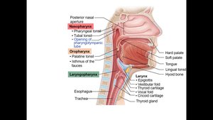
The pharynx is a muscular tube that serves as a passageway for both air and food. It is divided into three regions:
Nasopharynx: Contains pharyngeal tonsils (adenoids) and pharyngotympanic tubes for middle ear pressure equalization
Oropharynx: Passageway for food and air from the soft palate to the epiglottis
Laryngopharynx: Passageway for food and air, continuous with the esophagus
Clinical Note: Adenoid Hypertrophy
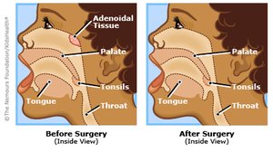
Enlarged or infected adenoids can block the nasopharynx, forcing mouth breathing and reducing air filtration, warming, and moistening. Chronic enlargement can disturb speech and sleep.
Lower Respiratory System
The Larynx
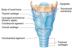
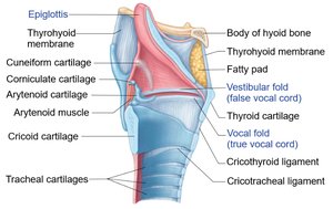
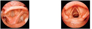
The larynx, or voice box, is responsible for maintaining an open airway, routing air and food into proper channels, and voice production. It contains the vocal folds (true vocal cords) and the glottis (the opening between the folds).
Vocal folds: Vibrate to produce sound as air passes through
Pitch: Determined by the length and tension of the vocal cords
Loudness: Determined by the force of the airstream
Sphincter Functions of the Larynx
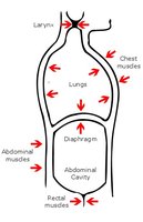
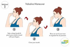
The vocal folds can act as a sphincter to prevent air passage, as seen in the Valsalva maneuver, which increases intra-abdominal pressure and stabilizes the trunk during heavy lifting.
Clinical Note: Laryngitis
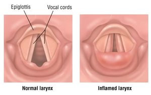
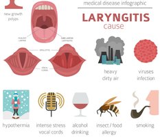
Laryngitis is the inflammation of the vocal folds, causing swelling and interfering with vibration. This leads to hoarseness or loss of voice and is commonly caused by viral infections, overuse, or irritants.
The Trachea
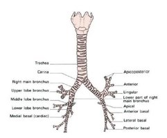
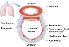
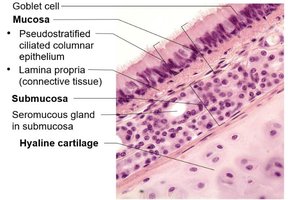
The trachea, or windpipe, extends from the larynx and divides into the right and left main bronchi. Its wall is composed of three layers: mucosa (ciliated pseudostratified epithelium), submucosa (with seromucous glands), and adventitia (outer connective tissue). C-shaped cartilage rings prevent collapse.
Clinical Note: Smoking and Tracheal Obstruction
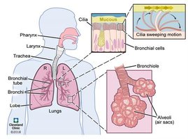
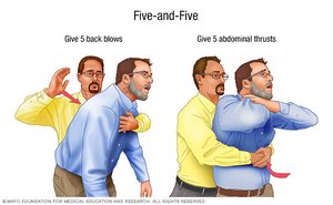
Smoking destroys cilia, making coughing the only way to clear mucus. Tracheal obstruction is life-threatening and may require the Heimlich maneuver to expel blockages.
The Bronchi and Bronchial Tree
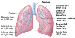
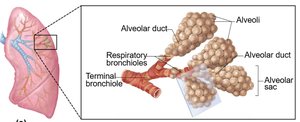
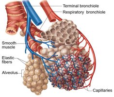
The trachea branches into the right and left main bronchi, which further divide into smaller bronchi and bronchioles, forming the bronchial tree. Bronchioles lead to terminal bronchioles, respiratory bronchioles, alveolar ducts, and alveolar sacs, where gas exchange occurs.
The Alveoli and Respiratory Membrane
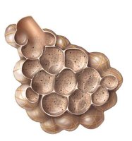
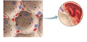
Alveoli are tiny air sacs where gas exchange occurs. The respiratory membrane is a thin barrier (~0.5 μm) composed of alveolar and capillary walls, allowing efficient diffusion of gases. Type I alveolar cells form the structure, while Type II cells secrete surfactant to reduce surface tension. Alveolar macrophages keep the surfaces sterile.
Lungs and Pleurae
Lung Anatomy
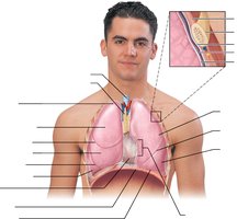
The lungs are divided into lobes (three on the right, two on the left) and contain the bronchial tree, alveoli, and associated blood vessels. The hilum is the entry/exit site for bronchi, blood vessels, lymphatics, and nerves.
Pleurae
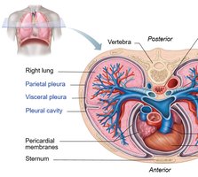
The pleurae are double-layered serous membranes surrounding each lung. The parietal pleura lines the thoracic wall, while the visceral pleura covers the lung surface. Pleural fluid in the pleural cavity reduces friction and assists lung expansion and recoil.
Clinical Note: Pleurisy and Pleural Effusion
Pleurisy is inflammation of the pleurae, often causing pain and friction. Excess fluid accumulation (pleural effusion) can hinder breathing.
Mechanics of Breathing (Pulmonary Ventilation)
Pressure Relationships
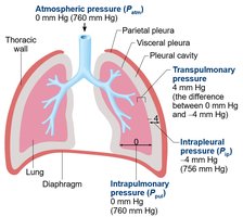
Breathing involves changes in thoracic volume and pressure. Key pressures include:
Atmospheric pressure (Patm): Pressure exerted by air around the body (760 mm Hg at sea level)
Intrapulmonary pressure (Ppul): Pressure in alveoli, fluctuates with breathing
Intrapleural pressure (Pip): Always negative relative to Patm and Ppul, helps keep lungs inflated
Pneumothorax
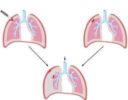
Pneumothorax is the presence of air in the pleural cavity, causing lung collapse due to loss of negative intrapleural pressure.
Inspiration and Expiration
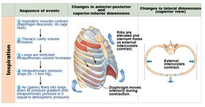
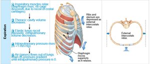
Inspiration is an active process involving the diaphragm and external intercostal muscles, increasing thoracic volume and decreasing pressure to draw air in. Expiration is usually passive, relying on muscle relaxation and lung recoil. Forced expiration uses abdominal and internal intercostal muscles.
Nonrespiratory Air Movements
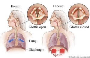
Movements such as coughing, sneezing, hiccups, and yawning modify normal respiratory rhythm, often as reflex actions.
Physical Factors Influencing Pulmonary Ventilation
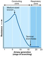
Airway resistance: Friction in airways; reduced by bronchodilation
Alveolar surface tension: Reduced by surfactant to prevent alveolar collapse
Lung compliance: The ease with which lungs expand; decreased by fibrosis or inflammation
Assessing Ventilation
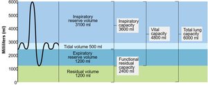
A spirometer measures respiratory volumes:
Tidal volume (TV): Air moved in/out with each breath
Inspiratory reserve volume (IRV): Extra air inhaled beyond TV
Expiratory reserve volume (ERV): Extra air exhaled beyond TV
Residual volume (RV): Air remaining in lungs after forced expiration
Gas Exchange
External and Internal Respiration
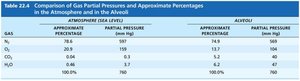
Gas exchange occurs by diffusion across the respiratory membrane (external respiration) and between blood and tissues (internal respiration). Governed by Dalton’s law of partial pressures, each gas moves down its partial pressure gradient.
Gas | Atmosphere (%) | Atmosphere (mm Hg) | Alveoli (%) | Alveoli (mm Hg) |
|---|---|---|---|---|
N2 | 78.6 | 597 | 74.9 | 569 |
O2 | 20.9 | 159 | 13.7 | 104 |
CO2 | 0.04 | 0.3 | 5.2 | 40 |
H2O | 0.46 | 3.7 | 6.2 | 47 |
Total | 100.0 | 760 | 100.0 | 760 |
Oxygen and Carbon Dioxide Transport
Oxygen: 1.5% dissolved in plasma, 98.5% bound to hemoglobin (Hb)
Carbon dioxide: 7–10% dissolved in plasma, 20% bound to Hb (carbaminohemoglobin), 70% as bicarbonate ions (HCO3-) in plasma
The chloride shift and carbonic anhydrase facilitate CO2 transport and conversion in red blood cells.
Control of Respiration
Neural and Chemical Regulation
Respiratory rhythms are regulated by the medullary and pontine respiratory centers, chemoreceptors (central and peripheral), and higher brain centers. Rising CO2 is the most powerful respiratory stimulant. Peripheral chemoreceptors in the carotid and aortic bodies respond to significant drops in O2 or pH.
Respiratory System Disorders
Chronic Obstructive Pulmonary Disease (COPD)
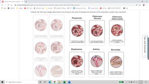
COPD includes chronic bronchitis and emphysema, characterized by irreversible airflow limitation, dyspnea, and frequent infections. Smoking is a major risk factor.
Asthma
Asthma is marked by acute episodes of airway inflammation, bronchospasm, and reversible airflow obstruction, often triggered by allergens or irritants.
Tuberculosis (TB)
TB is an infectious disease caused by Mycobacterium tuberculosis, leading to chronic cough, fever, and weight loss. Treatment requires prolonged antibiotic therapy.
Lung Cancer
Lung cancer is the leading cause of cancer death, with smoking as the primary risk factor. Types include adenocarcinoma, squamous cell carcinoma, and small cell carcinoma.
Sleep Apnea
Sleep apnea involves repeated cessation of breathing during sleep, leading to daytime sleepiness and increased risk of cardiovascular disease. Obstructive sleep apnea is most common and can be treated with CPAP devices.
Cystic Fibrosis
Cystic fibrosis is a genetic disorder causing thick, sticky mucus that clogs airways and leads to recurrent infections. It is caused by mutations in the CFTR gene affecting chloride transport.
