 Back
BackChapter 22: The Respiratory System – Structure, Function, and Gas Exchange
Study Guide - Smart Notes
 Tailored notes based on your materials, expanded with key definitions, examples, and context.
The Respiratory System: Overview and Functions
The respiratory system is essential for gas exchange, speech, olfaction, pH regulation, blood pressure control, and aiding venous return. It consists of organs and structures that facilitate the movement of air and the exchange of gases between the atmosphere and the bloodstream.
Gas Exchange: Oxygen (O2) is absorbed and carbon dioxide (CO2) is expelled between the air and blood.
Speech and Olfaction: The system enables vocalization and houses olfactory receptors for the sense of smell.
pH Regulation: By eliminating CO2, the respiratory system helps control blood pH.- CO2 is a biproduct of cellular respiration.
Blood Pressure Regulation: The lungs produce angiotensin II, a vasoconstrictor.
Venous Return: Breathing assists in pumping blood and lymph back to the heart.
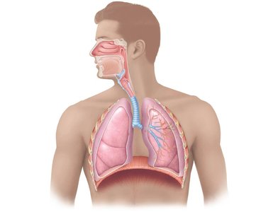
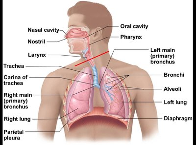
Major Structures of the Respiratory System
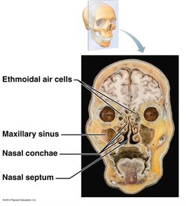
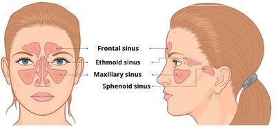
Nasal Cavity and Paranasal Sinuses
The nasal cavity provides an airway, moistens and warms air, filters particles, resonates sound for speech, and houses olfactory receptors. The paranasal sinuses open into the nasal cavity and are lined by respiratory mucosa, performing similar functions.
Turbinate Bones (Conchae): Increase surface area for warming and humidifying air.
Mucosa: Contains olfactory (smell) and respiratory (filter, heat, moisten) regions.
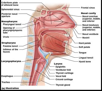
The Pharynx
The pharynx is a muscular tube that serves as a passageway for both air and food, divided into three regions:
Nasopharynx: Lined with pseudostratified ciliated columnar epithelium; contains the uvula and pharyngeal tonsil (adenoids).
Oropharynx: Located behind the mouth; lined with nonkeratinized stratified squamous epithelium; contains palatine and lingual tonsils.
Laryngopharynx: Passageway for food and air; continuous with the esophagus and larynx.
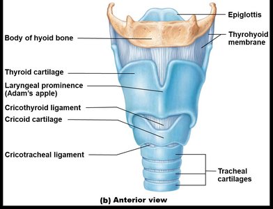
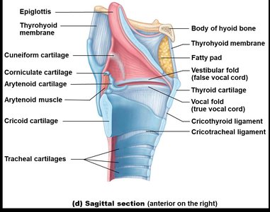
The Larynx
The larynx connects the pharynx to the trachea and is responsible for voice production, maintaining an open airway, and routing air and food using the epiglottis.
Cartilages: Includes thyroid, cricoid, arytenoid, and epiglottis.
Vocal Folds: True and false vocal cords for sound production and airway protection.
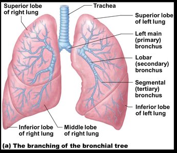
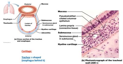
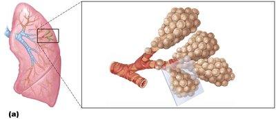
The Trachea and Bronchial Tree
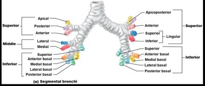
The trachea is a flexible tube supported by C-shaped cartilage rings, leading to the primary bronchi, which branch into secondary (lobar) and tertiary (segmental) bronchi, and finally into bronchioles and terminal bronchioles.
Trachea: Lined with pseudostratified ciliated columnar epithelium; supported by hyaline cartilage.
Bronchi: Right lung has three secondary bronchi, left lung has two; further branching forms the bronchial tree.
Bronchioles: Less than 1 mm in diameter; terminal bronchioles are less than 0.5 mm and lack cartilage.
Histology and Epithelial Types
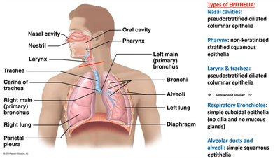
The respiratory tract is lined by different types of epithelia, which change along its length to suit functional needs.
Nasal cavity, larynx, trachea: Pseudostratified ciliated columnar epithelium.
Pharynx: Non-keratinized stratified squamous epithelium.
Bronchioles: Simple cuboidal epithelium (no cilia or mucous glands in respiratory bronchioles).
Alveoli: Simple squamous epithelium for gas exchange.
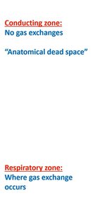
Conducting Zone vs. Respiratory Zone
The respiratory system is divided into the conducting zone (air passageways that filter, warm, and moisten air but do not participate in gas exchange) and the respiratory zone (where gas exchange occurs).
Conducting Zone: Includes nasal cavity, pharynx, larynx, trachea, bronchi, and terminal bronchioles.
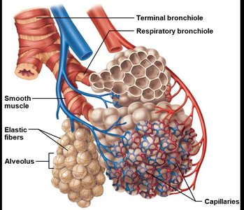
Respiratory Zone: Includes respiratory bronchioles, alveolar ducts, and alveoli.
Alveoli and Gas Exchange
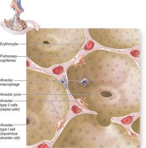
Structure of Alveoli
Alveoli are tiny air sacs where gas exchange occurs. They are surrounded by capillaries and have thin walls to facilitate diffusion.
Type I Alveolar Cells: Simple squamous cells forming the respiratory membrane.
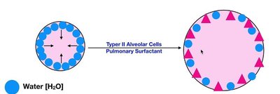
Type II Alveolar Cells: Cuboidal cells that secrete surfactant to reduce surface tension.
Alveolar Macrophages: Remove debris and pathogens.
Alveolar Pores: Equalize air pressure and provide alternate air routes.
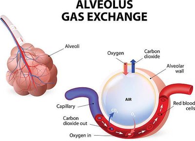
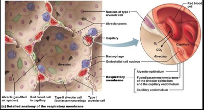
Gas Exchange Mechanism
Gas exchange occurs by diffusion across the respiratory membrane. Oxygen moves from alveoli into blood, while carbon dioxide moves from blood into alveoli to be exhaled.
Respiratory Membrane: Formed by alveolar and capillary walls and their fused basal laminae.
Surfactant: Reduces surface tension, preventing alveolar collapse.
Lungs and Pleurae
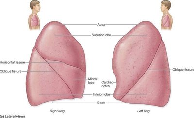
Lung Anatomy
The lungs are divided into lobes and segments, each supplied by its own bronchus and blood vessels. The right lung has three lobes; the left lung has two and a cardiac notch.
Bronchopulmonary Segments: Functionally independent units of the lung.
Pleurae: Double-layered serous membranes (parietal and visceral) that reduce friction and compartmentalize the lungs.
Respiratory Physiology and Terminology
Key Terms
Respiration: Overall process of gas exchange.
External Respiration: Gas exchange between air in the lungs and blood.
Internal Respiration: Gas exchange between blood and tissues.
Pulmonary Ventilation: Movement of air into (inspiration) and out of (expiration) the lungs.
Cellular Respiration: Metabolic processes (glycolysis, Krebs cycle, oxidative phosphorylation) that produce ATP.
CO2 Transport and Acid-Base Balance
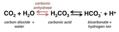
CO2 is transported in the blood as dissolved gas, carbaminohemoglobin, and bicarbonate. The following equation summarizes the conversion of CO2 to bicarbonate, which is crucial for acid-base balance:
Carbonic Anhydrase: Enzyme that catalyzes the reversible reaction between CO2 and water to form carbonic acid, which dissociates into bicarbonate and hydrogen ions.
pH Regulation: Removal of CO2 by the lungs helps maintain blood pH.
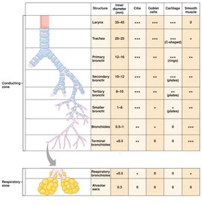
Summary Table: Conducting vs. Respiratory Zone Structures
Structure | Inner Diameter (mm) | Cilia | Goblet Cells | Cartilage | Smooth Muscle |
|---|---|---|---|---|---|
Larynx | 35–45 | +++ | +++ | +++ (plates) | + |
Trachea | 20–25 | +++ | +++ | +++ (C-shaped) | + |
Primary Bronchi | 12–16 | +++ | +++ | +++ (rings) | + |
Secondary Bronchi | 8–10 | +++ | +++ | ++ (plates) | ++ |
Tertiary Bronchi | 1–8 | +++ | +++ | + (plates) | +++ |
Smaller Bronchi | 1–8 | +++ | +++ | + (plates) | +++ |
Terminal Bronchioles | <0.5 | + | 0 | 0 | +++ |
Respiratory Bronchioles | <0.5 | 0 | 0 | 0 | ++ |
Alveolar Sacs | 0 | 0 | 0 | 0 | 0 |
Additional info: Table summarizes the structural and functional differences between the conducting and respiratory zones of the respiratory tract.
