 Back
BackChapter 23: The Digestive System – Anatomy & Physiology Study Guide
Study Guide - Smart Notes
 Tailored notes based on your materials, expanded with key definitions, examples, and context.
Overview of the Digestive System
Functions and Major Organs
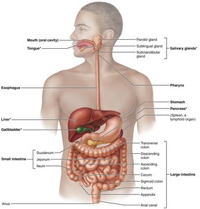
The digestive system is essential for converting food into nutrients that fuel and build the body. It consists of the alimentary canal (gastrointestinal tract) and accessory organs. The alimentary canal is a continuous muscular tube from mouth to anus, while accessory organs aid digestion by secreting fluids and enzymes.
Alimentary canal organs: Mouth, pharynx, esophagus, stomach, small intestine, large intestine, anus
Accessory organs: Teeth, tongue, gallbladder, salivary glands, liver, pancreas
Digestive Processes
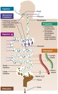
Six essential activities occur during digestion:
Ingestion: Intake of food
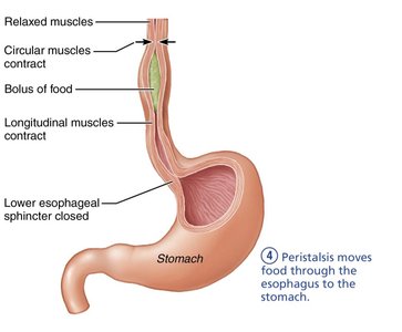
Propulsion: Movement of food via swallowing and peristalsis
Mechanical breakdown: Chewing, mixing, churning, and segmentation to increase surface area
Digestion: Enzymatic breakdown of food into absorbable molecules
Absorption: Passage of nutrients into blood or lymph
Defecation: Elimination of indigestible residues
Motility: Peristalsis and Segmentation
Peristalsis vs. Segmentation
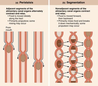
Motility in the GI tract is achieved by two main mechanisms:
Peristalsis: Alternating waves of contraction and relaxation propel food distally.
Segmentation: Nonadjacent segments contract and relax, mixing food and breaking it down mechanically.
Relationship to the Peritoneum
Peritoneal Structures
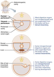
The peritoneum is a serous membrane lining the abdominopelvic cavity. It consists of:
Visceral peritoneum: Covers digestive organs
Parietal peritoneum: Lines the body wall
Peritoneal cavity: Space between membranes, lubricated by serous fluid
Mesentery: Double layer of peritoneum anchoring organs and providing routes for vessels and nerves
Organs are classified as intraperitoneal (suspended by mesentery) or retroperitoneal (behind the peritoneum).
Histology of the Alimentary Canal
Four Basic Layers (Tunics)
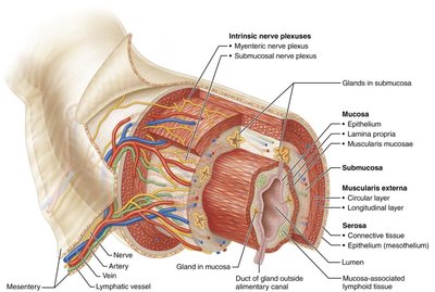
The alimentary canal wall consists of four layers:
Mucosa: Innermost layer; secretes mucus, enzymes, hormones; absorbs nutrients; protects against pathogens
Submucosa: Areolar connective tissue with blood vessels, lymphatics, nerve plexus, and elastic fibers
Muscularis externa: Smooth muscle responsible for peristalsis and segmentation; forms sphincters
Serosa: Outermost layer; visceral peritoneum; replaced by adventitia in retroperitoneal organs
The Mouth (Oral Cavity)
Anatomy and Functions
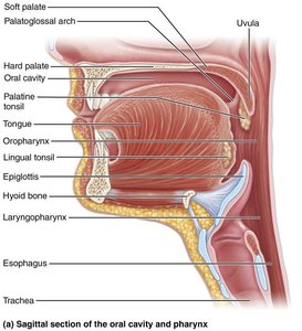
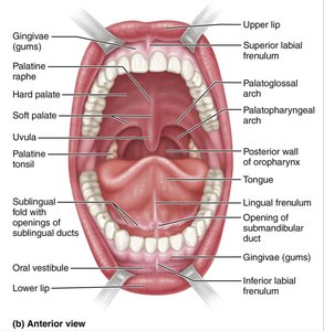
The mouth is the site of ingestion, mechanical breakdown, and the beginning of digestion. It is bounded by lips, cheeks, palate, and tongue, and lined with stratified squamous epithelium for protection.
Lips and cheeks: Aid in holding and manipulating food
Palate: Hard palate forms the roof; soft palate closes off nasopharynx during swallowing
Tongue: Mixes food, forms bolus, initiates swallowing, speech, and taste
The Tongue and Papillae
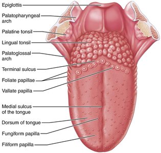
The tongue contains intrinsic and extrinsic muscles, and is attached by the lingual frenulum. Its surface bears papillae:
Filiform papillae: Provide friction, no taste buds
Fungiform papillae: Mushroom-shaped, contain taste buds
Vallate papillae: V-shaped row at back, contain taste buds
Foliate papillae: Lateral aspects, contain taste buds
The Salivary Glands
Types and Functions
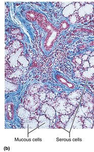
Salivary glands secrete saliva, which cleanses the mouth, dissolves food, moistens it, and begins starch digestion via amylase. Major glands include parotid, submandibular, and sublingual.
Serous cells: Produce watery, enzyme-rich secretion
Mucous cells: Produce mucus
The Teeth
Types and Structure
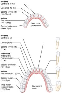
Teeth are essential for mastication (chewing), breaking food into smaller pieces. Humans have two sets:
Deciduous (primary) teeth: 20 baby teeth
Permanent teeth: 32 adult teeth, including wisdom teeth
Tooth Structure
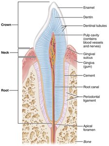
Each tooth has:
Crown: Exposed part, covered by enamel
Neck: Constricted region connecting crown and root
Root: Embedded in jawbone, covered by cementum
Dentin: Shock-absorbing, surrounds pulp cavity
Pulp cavity: Contains blood vessels and nerves
Periodontal ligament: Anchors tooth in socket
The Pharynx and Esophagus
Transport of Food
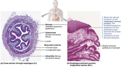
The pharynx and esophagus move food from the mouth to the stomach. The esophagus is a muscular tube with four tunics, including a mucosa of stratified squamous epithelium and an adventitia.
Swallowing (Deglutition)
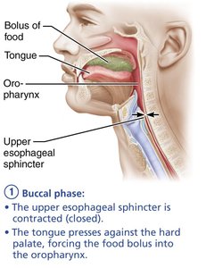
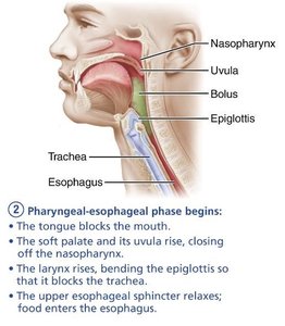
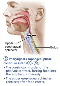
Swallowing involves coordinated muscle activity in two phases:
Buccal phase: Voluntary, tongue pushes bolus into oropharynx
Pharyngeal-esophageal phase: Involuntary, controlled by vagus nerve and swallowing center in brainstem
The Stomach
Gross Anatomy
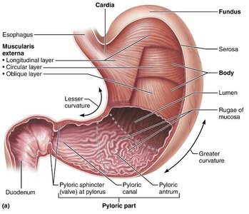
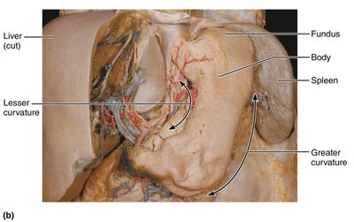
The stomach is a temporary storage tank, continuing physical and chemical breakdown of food. It has four main regions: cardia, fundus, body, and pyloric part. The stomach wall contains rugae (folds) for expansion.
Microscopic Anatomy
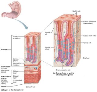
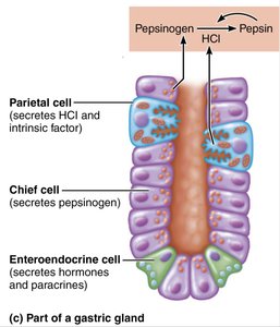
The stomach wall has three muscle layers and a mucosa with gastric pits leading to glands. Gland cells include:
Mucous neck cells: Secrete acidic mucus
Parietal cells: Secrete HCl and intrinsic factor
Chief cells: Secrete pepsinogen and gastric lipases
Enteroendocrine cells: Secrete hormones and paracrines
Clinical Note: Gastric Ulcers
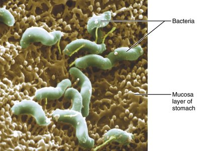
Most gastric ulcers are caused by Helicobacter pylori bacteria, which damage the mucosal barrier. Treatment includes antibiotics and acid-reducing drugs.
Regulation of Gastric Secretion and Motility
Phases of Gastric Secretion
Gastric secretion is regulated by neural and hormonal mechanisms, divided into three phases:
Cephalic phase: Triggered by sight, smell, taste, or thought of food
Gastric phase: Triggered by food entry, distension, and chemical stimuli
Intestinal phase: Brief stimulation followed by inhibition as chyme enters duodenum
Hormones and Paracrines in Digestion
Several hormones regulate digestive activities. The following table summarizes their sites of production, stimuli, target organs, and activities:
Hormone | Site of Production | Stimulus | Target Organ | Activity |
|---|---|---|---|---|
Cholecystokinin (CCK) | Duodenal mucosa | Fatty chyme | Stomach, pancreas, gallbladder | Inhibits stomach, increases pancreatic juice, contracts gallbladder |
Gastrin | Stomach mucosa | Food, ACh | Stomach, small intestine | Increases HCl, stimulates motility |
Secretin | Duodenal mucosa | Acidic chyme | Stomach, pancreas, liver | Inhibits gastric secretion, increases bile and bicarbonate |
Somatostatin | Stomach/duodenal mucosa | Food, sympathetic stimulation | Stomach, pancreas, intestine | Inhibits secretion and motility |
Vasoactive Intestinal Peptide (VIP) | Enteric neurons | Chyme | Small intestine, pancreas, stomach | Stimulates buffer secretion, increases blood flow, inhibits acid |
Mechanism of HCl Secretion
Parietal cells pump H+ into the stomach lumen via H+/K+ ATPases. Cl- diffuses in to form HCl. The process creates an alkaline tide as HCO3- is exported to blood.
The Liver, Gallbladder, and Pancreas
Liver Anatomy and Functions
The liver is the largest gland, producing bile for fat emulsification. It consists of four lobes and is anchored by ligaments. Blood enters via hepatic artery and portal vein; bile leaves via hepatic ducts.
Microscopic Anatomy of the Liver
Liver lobules are hexagonal units with plates of hepatocytes radiating from a central vein. Portal triads contain branches of hepatic artery, portal vein, and bile duct. Sinusoids are leaky capillaries lined with macrophages.
Bile Composition and Circulation
Bile contains bile salts, pigments (bilirubin), cholesterol, and phospholipids. Bile salts are recycled via enterohepatic circulation.
Gallbladder and Pancreas
The gallbladder stores and concentrates bile. The pancreas produces digestive enzymes and bicarbonate-rich juice, with both exocrine and endocrine functions.
Activation of Pancreatic Proteases
Proteases are secreted inactive and activated in the duodenum by enteropeptidase, which converts trypsinogen to trypsin.
Relationship to Duodenum
Bile and pancreatic juice enter the duodenum via regulated ducts.
The Small Intestine
Structure and Function
The small intestine is the major site for digestion and absorption. Its length and structural modifications (circular folds, villi, microvilli) greatly increase surface area.
Histology and Cell Types
Enterocytes: Absorptive cells with microvilli
Goblet cells: Secrete mucus
Enteroendocrine cells: Secrete hormones
Paneth cells: Secrete antimicrobial agents
Stem cells: Continuously divide and replace epithelium
The Large Intestine
Structure and Function
The large intestine absorbs water and eliminates feces. It includes the cecum, appendix, colon (ascending, transverse, descending, sigmoid), rectum, and anal canal. Unique features include teniae coli, haustra, and epiploic appendages.
Microscopic Anatomy
The large intestine is lined with simple columnar epithelium (except anal canal, which is stratified squamous). It lacks villi and brush border, but has deep crypts with many goblet cells for mucus secretion.
Digestive Processes
Haustral contractions: Slow segmenting movements for mixing
Mass movements: Powerful waves moving feces toward rectum
Defecation: Reflex triggered by rectal distension, involving voluntary and involuntary sphincters
Physiology of Digestion and Absorption
Enzymatic Hydrolysis
Digestion is a catabolic process breaking macromolecules into monomers via hydrolysis, primarily in the small intestine.
Mechanisms of Absorption
Most nutrients are absorbed through enterocytes by active or passive transport. Lipids are absorbed via simple diffusion and transported in chylomicrons.
Processing of Nutrients
Carbohydrates
Digestion begins in mouth (salivary amylase), continues in small intestine (pancreatic amylase, brush border enzymes)
Monosaccharides absorbed via secondary active transport with Na+
Proteins
Digestion begins in stomach (pepsin), continues in small intestine (pancreatic proteases, brush border enzymes)
Amino acids absorbed via secondary active transport with Na+
Lipids
Emulsified by bile salts, digested by pancreatic lipase
Absorbed as micelles, reassembled into triglycerides, transported as chylomicrons
Nucleic Acids
Digested by pancreatic nucleases and brush border enzymes
Absorbed as nitrogenous bases, pentose sugars, and phosphate ions
Absorption of Vitamins, Electrolytes, and Water
Fat-soluble vitamins absorbed with lipids; water-soluble vitamins by passive or active transport
Electrolytes actively transported; water absorbed by osmosis
Summary Table: Functions of GI Organs
Clinical Notes
Peritonitis: Inflammation of peritoneum, often from ruptured appendix
Xerostomia: Dry mouth due to reduced saliva
Dental caries: Cavities from bacterial action
Heartburn: Acid reflux into esophagus
Gastric ulcers: Often caused by H. pylori
Hepatitis and cirrhosis: Liver diseases
Gallstones: Crystallized cholesterol in gallbladder
Appendicitis: Inflammation of appendix
Diverticulosis: Herniations in colon wall
Malabsorption: Impaired nutrient absorption, e.g., celiac disease
