 Back
BackChapter 23: The Digestive System – Structure, Function, and Regulation
Study Guide - Smart Notes
 Tailored notes based on your materials, expanded with key definitions, examples, and context.
Chapter 23: The Digestive System
Overview of the Digestive System
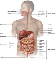
The digestive system is responsible for breaking down food into nutrients, which are then absorbed into the blood to fuel cellular processes. It consists of the alimentary canal (gastrointestinal tract) and accessory digestive organs. The GI tract is a continuous tube running from the mouth to the anus, while accessory organs assist in digestion through secretion and mechanical processing.
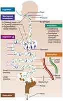
Major Processes of the Digestive System
Ingestion: Taking food into the mouth.
Propulsion: Moving food through the GI tract, including swallowing (voluntary) and peristalsis (involuntary waves of contraction and relaxation).
Mechanical Breakdown: Physically breaking food into smaller pieces to increase surface area for enzymes. Includes chewing, mixing, churning, and segmentation.
Digestion: Chemical breakdown of food into absorbable molecules by enzymes.
Absorption: Transport of digested nutrients from the GI tract lumen into blood or lymph.
Defecation: Elimination of indigestible substances as feces.
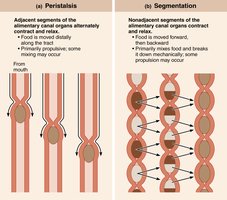
Peristalsis vs. Segmentation
Peristalsis: Propulsive movement; adjacent segments of the GI tract alternately contract and relax to move food distally.
Segmentation: Mechanical breakdown; nonadjacent segments contract and relax, mixing food and breaking it down mechanically.
23.2 The GI Tract Has Four Layers and Is Usually Surrounded by Peritoneum
Relationship of Digestive Organs to the Peritoneum
The peritoneum is a serous membrane lining the abdominopelvic cavity. It consists of:
Visceral peritoneum: Covers external surfaces of most digestive organs.
Parietal peritoneum: Lines the body wall.
Peritoneal cavity: Space between the two layers, filled with lubricating serous fluid.
Mesentery: Double layer of peritoneum that supports digestive organs, stores fat, and provides a route for blood vessels, lymphatics, and nerves.
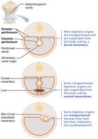
Organs are classified as:
Intraperitoneal: Surrounded by peritoneum and suspended by mesentery.
Retroperitoneal: Located posterior to the peritoneum (e.g., most of the pancreas, duodenum, parts of the large intestine).

Histology of the Alimentary Canal
The walls of the alimentary canal share four basic layers (tunics):
Mucosa: Innermost layer; secretes mucus, digestive enzymes, and hormones; absorbs nutrients; protects against pathogens.
Submucosa: Areolar connective tissue with blood and lymphatic vessels, lymphoid follicles, and nerve fibers; allows stretch and recoil.
Muscularis externa: Responsible for segmentation and peristalsis; consists of inner circular and outer longitudinal smooth muscle layers.
Serosa: Outermost layer (visceral peritoneum); areolar connective tissue covered with mesothelium. In retroperitoneal organs, the serosa is replaced by adventitia (dense connective tissue).
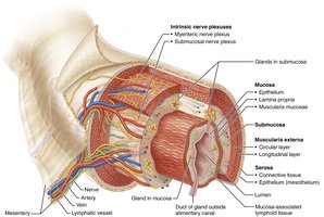
23.3 The GI Tract Nervous System: The Enteric Nervous System
The GI tract has its own nervous system, the enteric nervous system (ENS), sometimes called the "gut brain." It contains more neurons than the spinal cord and regulates digestive activity independently of the central nervous system (CNS).
Submucosal nerve plexus: Regulates glands and smooth muscle in the mucosa.
Myenteric nerve plexus: Controls GI tract motility; located between the circular and longitudinal muscle layers.
Neural Reflex Pathways
Short reflexes: Mediated by the ENS in response to stimuli within the GI tract.
Long reflexes: Involve the CNS and autonomic nerves, responding to stimuli inside or outside the GI tract.
Parasympathetic input: Stimulates digestive activity.
Sympathetic input: Inhibits digestive activity.
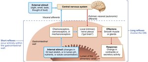
Regulation of Digestive Activity
Neural controls: Intrinsic (short reflexes) and extrinsic (long reflexes) regulate secretion and motility.
Hormonal controls: Hormones from the stomach and small intestine affect digestive organs.
Receptors: Mechanoreceptors and chemoreceptors in the GI tract wall respond to stretch, osmolarity, pH, and chemical composition.

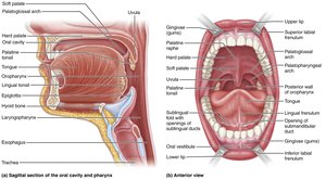
23.4 The Mouth: Ingestion and Initial Digestion
The mouth (oral or buccal cavity) is the entry point for food, where mechanical and chemical digestion begins. It is bounded by the lips, cheeks, palate, and tongue.
Tongue: Manipulates food, mixes it with saliva, and forms a bolus for swallowing.
Salivary glands: Secrete saliva to moisten food, begin starch digestion, and protect against microbes.
Teeth: Masticate (chew) food, breaking it into smaller pieces.

The Palate
Hard palate: Bony anterior roof of the mouth.
Soft palate: Muscular posterior roof; rises to close off the nasopharynx during swallowing.
Uvula: Fingerlike projection from the soft palate.
Salivary Glands
Parotid gland: Anterior to the ear; duct opens near the second upper molar.
Submandibular gland: Medial to the mandible; duct opens at the base of the tongue.
Sublingual gland: Under the tongue; multiple ducts open into the floor of the mouth.
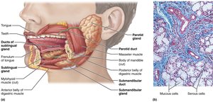
Saliva composition: Mostly water, slightly acidic, contains electrolytes, salivary amylase, lingual lipase, mucin, lysozyme, IgA, and metabolic wastes. Salivation is controlled by the parasympathetic nervous system and is stimulated by food, smell, or thought of food.
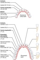
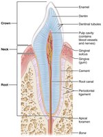
Teeth and Dentition
Deciduous teeth: 20 primary teeth, erupt between 6–24 months.
Permanent teeth: 32 teeth, replace primary teeth between ages 6–12 years.
Types: Incisors (cutting), canines (tearing), premolars and molars (grinding).
Digestive Processes in the Mouth
Ingestion
Mechanical breakdown: Mastication (chewing)
Propulsion: Initiation of swallowing (buccal phase)
Chemical digestion: Salivary amylase begins starch breakdown
23.5 The Pharynx and Esophagus: Food Conduction
The pharynx and esophagus serve as passageways for food from the mouth to the stomach. Their main function is propulsion via swallowing (deglutition).
Pharynx: Oropharynx and laryngopharynx allow passage of food, fluids, and air; lined with stratified squamous epithelium.
Esophagus: Muscular tube that propels food to the stomach by peristalsis.
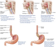
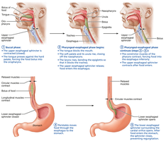
Swallowing (Deglutition)
Buccal phase: Voluntary; tongue pushes bolus into the pharynx.
Pharyngeal-esophageal phase: Involuntary; controlled by the swallowing center in the brainstem, involves peristalsis and closure of the nasopharynx and airway.

23.6 The Stomach: Temporary Storage and Protein Digestion
The stomach acts as a temporary storage tank, continues mechanical and chemical digestion, and delivers chyme to the small intestine. It produces gastric juice, churns food, and begins protein digestion.
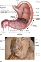
Regions: Cardia, fundus, body, pyloric part (antrum, canal, pylorus)
Curvatures: Greater (lateral) and lesser (medial)
Mesenteries: Lesser omentum (to liver), greater omentum (drapes over intestines)
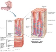
Microscopic Anatomy of the Stomach
Muscularis externa: Has an additional oblique layer for enhanced churning and mixing.
Mucosa: Simple columnar epithelium with mucous cells; contains gastric pits leading to gastric glands.

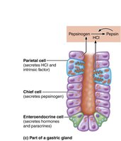
Gastric Gland Cells
Mucous neck cells: Secrete thin, acidic mucus.
Parietal cells: Secrete hydrochloric acid (HCl) and intrinsic factor (for vitamin B12 absorption).
Chief cells: Secrete pepsinogen (activated to pepsin by HCl) and gastric lipase.
Enteroendocrine cells: Secrete hormones and paracrines (e.g., gastrin, histamine, serotonin, somatostatin).
Mucosal Barrier
Protects stomach from self-digestion by acid and enzymes.
Consists of a thick bicarbonate-rich mucus, tight junctions between epithelial cells, and rapid cell turnover.
Digestive Processes in the Stomach
Propulsion: Peristaltic waves move food toward the pylorus.
Mechanical breakdown: Churning action mixes food with gastric juice.
Chemical digestion: HCl denatures proteins; pepsin digests proteins; gastric lipase digests fats (minor role).
Absorption: Limited to lipid-soluble substances (e.g., alcohol, aspirin).
Essential function: Secretion of intrinsic factor for vitamin B12 absorption (required for red blood cell maturation).
Regulation of Gastric Secretion and Motility
Neural mechanisms: Vagus nerve (parasympathetic) stimulates secretion; sympathetic input inhibits.
Hormonal mechanisms: Gastrin stimulates HCl secretion; enterogastrones from the small intestine inhibit gastric activity.
Phases of gastric secretion: Cephalic (before food enters), gastric (food in stomach), and intestinal (chyme in duodenum).
Gastric Motility and Emptying
Stomach stretches to accommodate food (receptive relaxation and gastric accommodation).
Peristaltic waves: Most vigorous in the pyloric region; only small amounts of chyme enter the duodenum at a time.
Regulation: Duodenal receptors control gastric emptying via enterogastric reflex and hormones (enterogastrones).
Example: A high-fat meal delays gastric emptying, while carbohydrate-rich chyme moves quickly into the duodenum.
Additional info: This guide covers the structure, function, and regulation of the digestive system from ingestion to the stomach, as outlined in Chapter 23 of Marieb's Human Anatomy & Physiology. It is suitable for ANP college students preparing for exams on the digestive system.
