 Back
BackChapter 23: The Respiratory System – Structure, Function, and Regulation
Study Guide - Smart Notes
 Tailored notes based on your materials, expanded with key definitions, examples, and context.
The Respiratory System: Overview
Introduction to the Respiratory System
The respiratory system is essential for gas exchange, supplying oxygen to tissues and removing carbon dioxide produced by cellular metabolism. It consists of specialized structures that facilitate the movement and exchange of gases between the external environment and the bloodstream.
Key Functions: Oxygen uptake, carbon dioxide removal, and protection of delicate exchange surfaces.
Gas Transport: Blood carries oxygen from lungs to tissues and returns carbon dioxide for exhalation.
Organization and Anatomy of the Respiratory System
Upper and Lower Respiratory Tracts
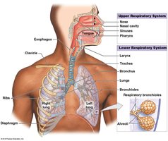
The respiratory system is divided into upper and lower tracts, each with distinct anatomical features and functions.
Upper Respiratory Tract: Nose, nasal cavity, sinuses, pharynx – filters, warms, and humidifies air.
Lower Respiratory Tract: Larynx, trachea, bronchi, bronchioles, alveoli – conducts air and facilitates gas exchange.
Respiratory Mucosa and Defense Mechanisms
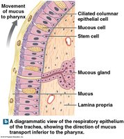
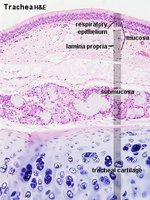
The conducting portions are lined with respiratory mucosa, consisting of an epithelium and lamina propria. This mucosa is equipped with defense mechanisms to filter and protect against pathogens and debris.
Mucous Glands: Produce mucus to trap particles.
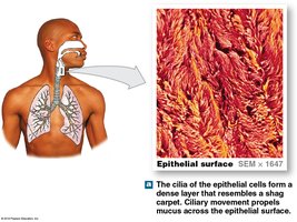
Cilia: Propel mucus toward the pharynx for removal.
Alveolar Macrophages: Engulf small particles reaching the alveoli.

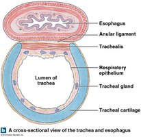
Microscopic Structure of the Respiratory Tract
Respiratory Epithelium
The respiratory epithelium is primarily pseudostratified ciliated columnar epithelium, with goblet cells producing mucus.
Lamina Propria: Areolar tissue supporting the epithelium.
Submucosa: Contains glands and connective tissue.
Tracheal Cartilage: Maintains airway patency.
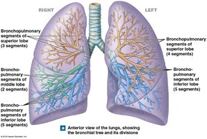
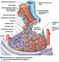
Airway Structure and Branching
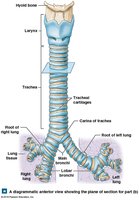
Trachea and Bronchial Tree
The trachea divides into right and left bronchi, which further branch into bronchioles and terminal bronchioles, forming the bronchial tree.
Bronchi: Supported by cartilage plates.
Bronchioles: Lack cartilage, dominated by smooth muscle.
Terminal Bronchioles: Lead to alveolar ducts and sacs.
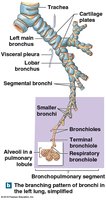
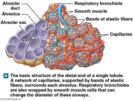
Alveolar Structure and Gas Exchange
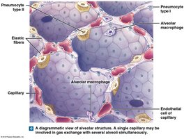
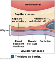
Alveoli and Blood Air Barrier
Alveoli are the primary sites of gas exchange, surrounded by capillaries and elastic fibers. The blood air barrier consists of three layers: alveolar cell layer, capillary endothelium, and fused basement membrane.
Pneumocytes Type I: Thin cells for gas exchange.
Pneumocytes Type II: Produce surfactant to reduce surface tension.
Alveolar Macrophages: Patrol and remove debris.
Pleura and Lung Cavities
Pleural Membranes
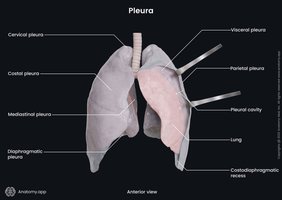
Each lung is contained within a pleural cavity lined by serous membranes.
Parietal Pleura: Lines thoracic wall.
Visceral Pleura: Covers lung surface.
Pleural Fluid: Lubricates space between layers, reducing friction.
Respiratory Physiology: External and Internal Respiration
Processes of Respiration
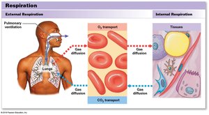
Respiration involves external and internal processes:
External Respiration: Exchange of O2 and CO2 between lungs and blood.
Internal Respiration: Exchange of O2 and CO2 between blood and tissues.
Pulmonary Ventilation: Mechanics and Regulation
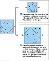
Boyle’s Law and Breathing Mechanics
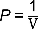
Pulmonary ventilation is governed by Boyle’s Law, which states that pressure and volume are inversely related in a closed system.
Equation:
Airflow: Air moves from high to low pressure.
Respiratory Cycle: Consists of inspiration (active) and expiration (passive or active).

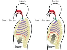
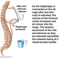
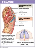
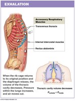
Muscles of Respiration
Inhalation: Diaphragm (75%), external intercostals (25%), accessory muscles (sternocleidomastoid, scalenes, pectoralis minor, serratus anterior).
Exhalation: Internal intercostals, transversus thoracis, abdominal muscles.
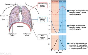
Pressure Changes and Lung Volumes
Intrapulmonary Pressure: Changes during breathing, determines airflow direction.
Intrapleural Pressure: Always below atmospheric, assists venous return.
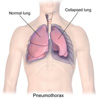
Pneumothorax: Air in pleural cavity causes lung collapse (atelectasis).
Respiratory Volumes and Capacities
Pulmonary Volumes
Tidal Volume (VT): Air moved in one breath.
Expiratory Reserve Volume (ERV): Air exhaled after tidal volume.
Residual Volume: Air remaining after maximal exhalation.
Inspiratory Reserve Volume (IRV): Air inhaled after tidal volume.
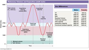
Respiratory Capacities
Inspiratory Capacity: VT + IRV
Functional Residual Capacity (FRC): ERV + Residual Volume
Vital Capacity: ERV + VT + IRV
Total Lung Capacity: Vital Capacity + Residual Volume

Volume/Capacity | Males (mL) | Females (mL) |
|---|---|---|
Tidal Volume (VT) | 500 | 500 |
Expiratory Reserve Volume (ERV) | 1000 | 700 |
Residual Volume | 1200 | 1100 |
Total Lung Capacity | 6000 | 4200 |
Functional Residual Capacity | 2200 | 1800 |
Gas Exchange: Physical Principles
Partial Pressures and Diffusion
Gas exchange depends on partial pressures and solubility.
Dalton’s Law: Total pressure is sum of partial pressures.
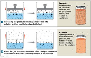
Henry’s Law: Gas solubility in liquid is proportional to partial pressure.
Efficiency: Short diffusion distance, large surface area, lipid solubility of O2 and CO2.
Transport of Gases in Blood
Oxygen Transport and Hemoglobin
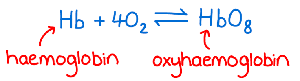
Oxygen is transported by binding to hemoglobin in red blood cells, forming oxyhemoglobin.
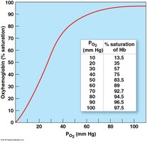
Hemoglobin Sat77uration: Percentage of heme units with bound O2.
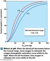
Factors Affecting Saturation: PO2, pH, temperature, metabolic activity.
Carbon Dioxide Transport
Carbon dioxide is carried in blood by three mechanisms:
Converted to carbonic acid (H2CO3).
Bound to hemoglobin.
Dissolved in plasma.
Control of Respiration
Neural Regulation
Respiratory rate and depth are controlled by centers in the brainstem (medulla oblongata and pons).
Dorsal Respiratory Group (DRG): Controls quiet breathing.
Ventral Respiratory Group (VRG): Controls forced breathing.
Apneustic and Pneumotaxic Centers: Adjust depth and rate.
Reflex Regulation
Chemoreceptors: Respond to changes in PCO2, PO2, and pH.
Baroreceptors: Respond to blood pressure changes.
Stretch Receptors: Respond to lung volume changes.
Age-Related Changes and Integration
Effects of Aging
Elastic tissue deteriorates, reducing compliance and vital capacity.
Arthritic changes restrict chest movement.
Emphysema increases with exposure to irritants.
Integration with Other Systems
The respiratory system works closely with the cardiovascular system to maintain homeostasis of O2 and CO2 levels in tissues.
Coordination is essential for efficient gas transport and exchange.
