 Back
BackChapter 9: The Muscular System – Structure, Function, and Major Muscles
Study Guide - Smart Notes
 Tailored notes based on your materials, expanded with key definitions, examples, and context.
The Muscular System
Introduction
The muscular system is essential for movement, posture, and many physiological processes. Skeletal muscles, the primary focus of this chapter, are voluntary muscles responsible for body movements, heat generation, and stabilization of joints. Understanding their structure, function, and organization is fundamental for students of anatomy and physiology.
Structure of Skeletal Muscles
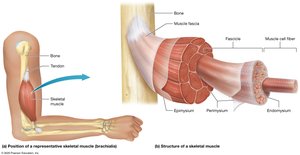
Gross Anatomy of a Skeletal Muscle
Skeletal Muscle Fibers: Long, thin cells surrounded by a thin layer of extracellular matrix called the endomysium.
Fascicle: A bundle of 10–100 muscle fibers, surrounded by perimysium.
Epimysium: Connective tissue that surrounds all fascicles in a muscle.
Fascia: The most superficial connective tissue sheath, continuous with the epimysium.
Tendons: Attach muscle to bone or other structures.
Rich Blood and Nerve Supply: Necessary for muscle function and voluntary control.
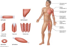
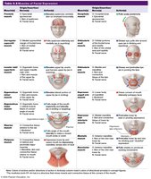
Fascicle Patterns and Muscle Shapes
Parallel: Evenly spaced fascicles; e.g., Sartorius.
Convergent: Broad at one end, tapers to a single tendon; e.g., Pectoralis Major.
Circular (Sphincters): Encircle openings; e.g., Orbicularis Oculi.
Fusiform: Thick in the middle, tapered at ends; e.g., Biceps Brachii.
Pennate: Fascicles attach at an angle to the tendon (uni-, bi-, or multipennate); e.g., Deltoid.
Naming Muscles
Muscles are named based on size (major, minor, longus, brevis, vastus), location (superior, inferior, medial, lateral), shape, appearance, function (flexor, extensor, adductor, abductor), and attachments (e.g., Sternocleidomastoid).
Names often provide clues to muscle action or position.
Functions of Skeletal Muscles
Muscle Tension and Movement
Muscle Tension: Force generated by muscle contraction, enabling movement and heat production.
Heat Generation: Conversion of ATP chemical energy to mechanical energy produces heat (e.g., shivering).
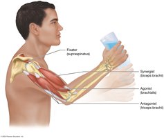
Functional Groups of Muscles
Agonist (Prime Mover): Main muscle responsible for movement.
Antagonist: Opposes or slows the action of the agonist.
Synergist: Assists the agonist for smooth movement.
Fixator: Stabilizes a bone or joint during movement.
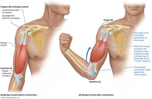
Muscle Origin and Insertion
Origin (O): More fixed attachment point.
Insertion (I): More moveable attachment point.
Muscle contraction typically moves the insertion toward the origin.
Example: Biceps brachii—origin at scapula, insertion at radius.
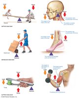
Lever Systems in Body Movements
Muscles and bones form lever systems to move loads around joints (fulcrums):
First-Class Lever: Fulcrum between load and force (e.g., atlanto-occipital joint).
Second-Class Lever: Load between fulcrum and force (e.g., metatarsophalangeal joints).
Third-Class Lever: Force between load and fulcrum (e.g., elbow joint with biceps brachii).
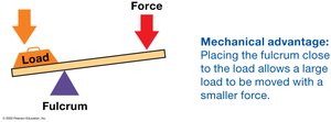
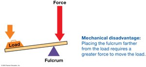
Mechanical Advantage and Disadvantage
Mechanical Advantage: Small force moves a large load (fulcrum close to load).
Mechanical Disadvantage: Greater force required to move the load (fulcrum farther from load).
Muscle Strains
Occurs when a muscle is overstretched or overexerted, causing tears.
Symptoms: Pain, swelling, bruising, limited movement.
Treatment: PRICE (Protect, Rest, Ice, Compression, Elevation), medications, physical therapy, or surgery if severe.

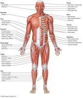
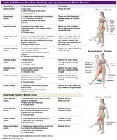
Studying Muscles: Overview of Major Muscle Groups
Muscles are categorized by region: head, neck, vertebral column, trunk, pelvic floor, pectoral girdle, upper limb, hip, and lower limb.
Familiarity with superficial anterior and posterior muscles is foundational for further study.

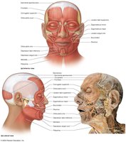
Muscles of Facial Expression
Key Muscles and Actions
Epicranius: Elevates eyebrows and forehead skin.
Orbicularis Oculi: Closes eyelids (blinking, winking).
Levator Palpebrae Superioris: Opens eyes.
Zygomaticus Major/Minor: Smiling.
Risorius: Lateral movement of mouth (smirk).
Orbicularis Oris: Controls lips for speech, eating, whistling.
Buccinator: Compresses cheek (sucking, chewing).
Platysma: Tenses neck skin, depresses jaw.
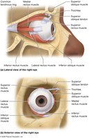
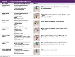
Extrinsic Eye Muscles
Muscles and Actions
Medial Rectus: Moves eye medially.
Lateral Rectus: Moves eye laterally.
Superior Rectus: Moves eye superiorly and medially.
Inferior Rectus: Moves eye inferiorly and medially.
Superior Oblique: Rotates eye inferiorly and laterally.
Inferior Oblique: Rotates eye superiorly and laterally.
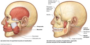
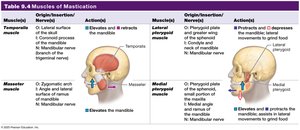
Muscles of Mastication and Swallowing
Muscles of Mastication
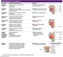
Masseter, Temporalis: Elevate mandible (close jaw).
Medial Pterygoid: Elevates mandible, assists in grinding.
Lateral Pterygoid: Depresses and protracts mandible, side-to-side movement.
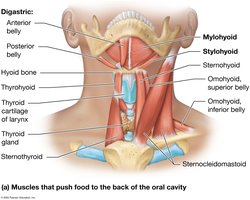
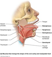
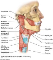
Muscles of Swallowing
Digastric, Stylohyoid, Mylohyoid, Geniohyoid: Elevate hyoid, raise tongue/floor of mouth.
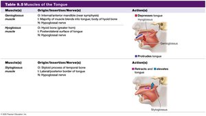
Genioglossus, Hyoglossus, Styloglossus: Move tongue for swallowing and speech.
Pharyngeal Constrictors: Push food into esophagus.
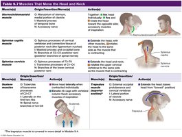
Muscles That Move the Head, Neck, and Vertebral Column
Major Muscles
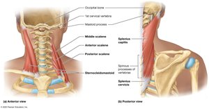
Sternocleidomastoid: Rotates and flexes head.
Scalenes: Lateral flexion of head, elevate ribs during deep breathing.
Trapezius: Extends head, moves scapula.
Splenius Capitis/Cervicis: Rotate/extend head and neck.
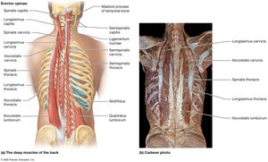
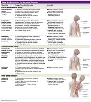
Muscles of the Vertebral Column
Erector Spinae Group: Extension and lateral flexion of vertebral column.
Transversospinal Group: Extension and rotation of vertebral column.
Quadratus Lumborum: Extension and lateral flexion of lumbar region.
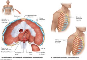
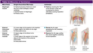
Muscles of Ventilation
Key Muscles
Diaphragm: Main muscle of inspiration; contracts and flattens to increase thoracic volume.
External Intercostals: Elevate ribs for inspiration.
Internal Intercostals: Depress ribs for forced expiration.
Sternocleidomastoid and Scalenes: Assist in forced inspiration.
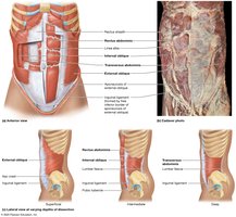
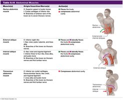
Abdominal Muscles
Major Muscles and Functions
Rectus Abdominis: Flexes trunk, increases intra-abdominal pressure.
External/Internal Obliques: Rotate and laterally flex trunk.
Transversus Abdominis: Compresses abdominal cavity.
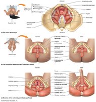
Muscles of the Pelvic Diaphragm, Urogenital Diaphragm, and Perineum
Pelvic Floor and Perineum
Levator Ani: Supports pelvic organs, divided into pubococcygeus, iliococcygeus, ischiococcygeus.
Urogenital Diaphragm: Contains external urethral sphincter (urination control), deep transverse perineal muscle (organ support).
External Anal Sphincter: Voluntary control of defecation.
Bulbospongiosus/Ischiocavernosus: Aid in erection and expulsion of semen (males).
Urinary Incontinence and Kegel Exercises
Weak pelvic floor muscles can cause urinary incontinence, especially after childbirth or with aging.
Kegel Exercises: Strengthen the levator ani muscle, help treat incontinence, and prevent pelvic organ prolapse.


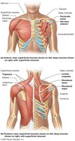
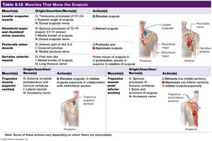
Muscles That Move the Scapula at the Pectoral Girdle
Major Muscles
Serratus Anterior: Protracts and rotates scapula.
Pectoralis Minor: Protracts and depresses scapula.
Trapezius: Elevates, retracts, depresses, and rotates scapula.
Levator Scapulae: Elevates scapula.
Rhomboid Major/Minor: Retract scapula.
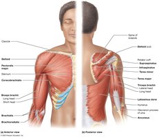
Muscles That Move the Arm at the Shoulder Joint
Major Muscles
Pectoralis Major: Flexes, adducts, and internally rotates arm.
Coracobrachialis: Assists with flexion and adduction.
Deltoid: Abducts, flexes, and extends arm.
Latissimus Dorsi: Extends, adducts, and internally rotates arm.
Teres Major: Assists latissimus dorsi.
Rotator Cuff Muscles: Stabilize shoulder joint (supraspinatus, infraspinatus, teres minor, subscapularis).
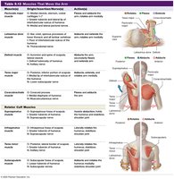
Rotator Cuff Injuries
Common in activities with repetitive overhead movements.
Symptoms: Pain, weakness, decreased range of motion.
Treatment: Rest, medications, physical therapy, or surgery.


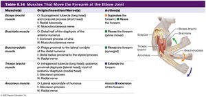
Muscles That Move the Forearm and Hand
Muscles at the Elbow Joint
Biceps Brachii: Flexes and supinates forearm.
Brachialis: Prime mover of elbow flexion.
Brachioradialis: Assists in flexion.
Triceps Brachii: Extends forearm.
Anconeus: Assists triceps in extension.
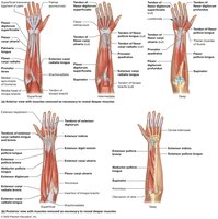
Muscles That Move the Hand and Fingers
Flexors: Anterior/medial forearm; flex hand and fingers.
Extensors: Posterior/lateral forearm; extend hand and fingers.
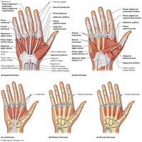
Intrinsic Hand Muscles: Fine movements of fingers and thumb.
Muscles of the Hip, Thigh, Knee, and Leg
Anterior and Medial Muscles
Iliopsoas: Thigh flexion.
Pectineus, Adductor Group, Gracilis: Thigh adduction.
Sartorius: Flexion, abduction, lateral rotation of thigh; flexion of leg.
Quadriceps Femoris: Extension of leg at knee; thigh flexion.
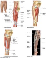

Posterior Muscles
Gluteal Group: Extension, abduction, rotation of thigh.
Hamstrings: Extension of thigh and leg.
Muscles of the Ankle, Foot, and Toes
Key Muscles and Actions
Tibialis Anterior, Extensor Digitorum Longus: Dorsiflexion of foot.
Fibularis Longus/Brevis: Eversion of foot.
Gastrocnemius, Soleus: Plantarflexion of foot.
Flexor Hallucis Longus, Flexor Digitorum Longus: Flex toes.
Intrinsic Foot Muscles: Support arches, fine movements.
The Big Picture of Muscle Movement
Additional info: This summary covers the structure, function, and organization of skeletal muscles, including their roles in movement, posture, and heat generation. It also provides an overview of major muscle groups and their actions, with relevant clinical correlations such as muscle strains, rotator cuff injuries, and the importance of pelvic floor exercises.
