 Back
BackChapter 9: The Muscular System – Structured Study Notes
Study Guide - Smart Notes
 Tailored notes based on your materials, expanded with key definitions, examples, and context.
The Muscular System
Structure of Skeletal Muscles
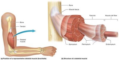
Skeletal muscles are composed of specialized cells called skeletal muscle fibers, which are long and thin. These fibers are surrounded by a thin layer of extracellular matrix known as the endomysium. Groups of 10–100 muscle fibers form a fascicle, surrounded by perimysium. All fascicles in a muscle are enclosed by epimysium, which is continuous with the fascia, the most superficial connective tissue sheath. Tendons attach muscles to bones or other structures, enabling movement. Skeletal muscles are richly supplied with blood vessels and nerves, and are voluntary, requiring nervous system stimulation to contract.
Fascicle Patterns and Muscle Shapes
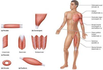
Muscles are classified by the arrangement of their fascicles and overall shape:
Parallel: Evenly spaced fascicles, tendon same width as muscle (e.g., Sartorius).
Convergent: Broad at one end, tapers to a single tendon (e.g., Pectoralis Major).
Circular: Encircles a structure, acts as a sphincter (e.g., Orbicularis Oculi).
Fusiform: Thick in the middle, tapered at ends (e.g., Biceps Brachii).
Pennate: Fascicles attach to tendon at an angle, feather-like appearance.
Unipennate: Fascicles on one side (e.g., Flexor Pollicis Longus).
Bipennate: Fascicles on both sides (e.g., Rectus Femoris).
Multipennate: Several tendons, fascicles attach like multiple feathers (e.g., Deltoid).
Naming Muscles
Muscles are named based on size, location, shape, appearance, position, function, and structural features:
Size: Major, Minor, Longus, Brevis, Vastus
Location: Superior, Inferior, Medial, Lateral, Anterior, Posterior, Profundus, Superficialis, Supra-, Infra-, External, Internal, Intercostal
Action: Flexor, Extensor, Levator, Adductor, Abductor, Pronator, Supinator, Depressor, Erector
Body Region: Abdominis, Brachii, Capitis, Carpi, Cervicis, Digitorum/Digiti, Femoris, Gluteal, Hallucis, Oculi, Oris, Pectoralis, Pollicis
Fiber Orientation: Oblique, Orbicular, Rectus, Transversus
Heads: Biceps (two), Triceps (three), Quadriceps (four)
Shape: Deltoid (triangular), Maximus (largest), Minimus/Minimi (smallest), Minor (small), Quadratus (rectangle), Rhomboid (rhombus), Serratus (jagged), Trapezius (trapezoid)
Functions of Skeletal Muscles
The primary function of skeletal muscles is to contract and generate muscle tension, which produces body movements and actions. Muscle tension also generates heat, converting chemical energy (ATP) into mechanical energy, with heat as a byproduct (e.g., shivering).
Functional Groups of Muscles
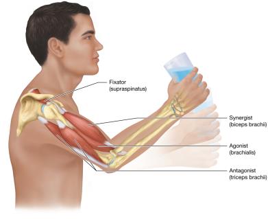
Agonists (Prime Movers): Provide most force for movement.
Antagonists: Oppose and slow the action, usually on the opposite side of the joint.
Synergists: Work with agonists to guide and smooth movement.
Fixators: Hold bones in place, making movement efficient and reducing injury risk.
Muscle Origin and Insertion
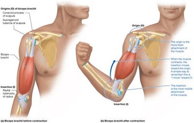
Origin (O): More fixed attachment point.
Insertion (I): Attachment point that moves.
Example: Biceps brachii attaches to scapula (origin) and radius (insertion).
Origin and insertion can switch depending on movement (e.g., Iliopsoas).
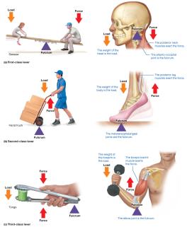
Lever Systems in Body Movements
Body movements use lever systems with four components:
Lever: Bone
Load: Object or body part being moved
Force: Muscle tension
Fulcrum: Joint
Types of levers:
First-Class Lever: Fulcrum in the middle (e.g., atlanto-occipital joint).
Second-Class Lever: Fulcrum at one end, load in the middle (e.g., metatarsophalangeal joints).
Third-Class Lever: Load at one end, force in the middle, fulcrum at the other end (e.g., elbow joint).
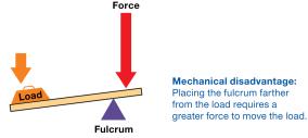
Mechanical Advantage and Disadvantage
Mechanical Advantage: Small force moves a large load.
Mechanical Disadvantage: Greater force required to move the load.
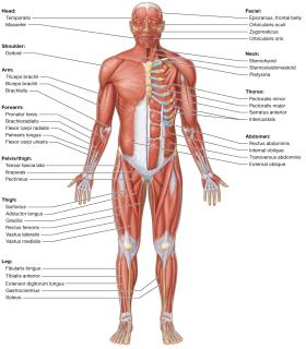
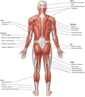
Studying Muscles: Superficial Muscles
Muscles are categorized by region: head, neck, vertebral column, trunk, pelvic floor, pectoral girdle, upper limb, hip, and lower limb. Familiarity with superficial muscles aids in understanding muscle locations and functions.
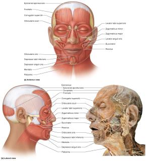
Muscles of Facial Expression
Overview
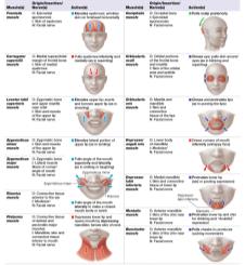
Facial muscles often insert into skin or connective tissue, producing shape changes in the face. Key muscles include:
Epicranius: Frontalis and Occipitalis, connected by epicranial aponeurosis; elevates eyebrows and forehead.
Orbicularis Oculi: Closes eyelids.
Levator Palpebrae Superioris: Opens eyelids.
Corrugator Supercilii: Squinting.
Levator Labii Superioris: Raises upper lip.
Zygomaticus Major/Minor: Smiling.
Risorius: Smirk.
Orbicularis Oris: Controls lips for speech, eating, whistling.
Depressor Anguli Oris, Depressor Labii Inferioris, Mentalis: Frowning, pouting.
Buccinator: Sucking, chewing, whistling.
Platysma: Jaw-dropping, tightens neck skin.
Extrinsic Eye Muscles
Overview
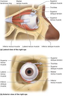
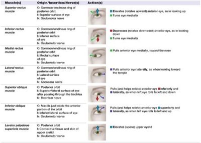
Six muscles move each eye, most originating from the common tendinous ring at the posterior orbit:
Medial Rectus: Moves eye medially.
Lateral Rectus: Moves eye laterally.
Superior Rectus: Moves eye superiorly and medially.
Inferior Rectus: Moves eye inferiorly and medially.
Superior Oblique: Rotates eye inferiorly and laterally via the trochlea.
Inferior Oblique: Rotates eye superiorly and laterally.
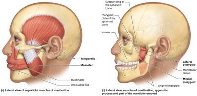
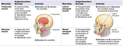
Muscles of Mastication
Overview
Muscles involved in chewing:
Masseter: Elevates mandible.
Temporalis: Elevates mandible.
Medial Pterygoid: Elevates mandible, synergist.
Lateral Pterygoid: Depresses mandible, protraction, side-to-side movement.
Muscles of Swallowing
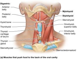
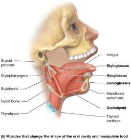
Pushing Food to the Posterior Oral Cavity
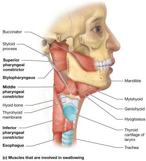
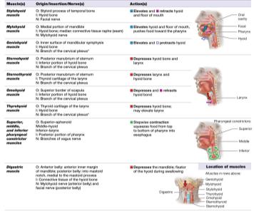
Digastric, Stylohyoid, Mylohyoid, Geniohyoid: Elevate hyoid bone, raise tongue and floor of mouth.
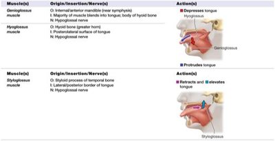
Genioglossus: Protrudes tongue.
Hyoglossus: Depresses tongue.
Styloglossus: Elevates and retracts tongue.
Pushing Food into the Esophagus
Sternohyoid, Omohyoid, Sternothyroid, Thyrohyoid: Depress pharynx, push food into esophagus.
Pharyngeal Constrictor: Contracts sequentially to push food into esophagus.
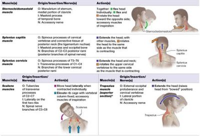
Muscles That Move the Head and Neck
Overview
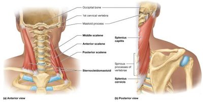
Sternocleidomastoid: Rotates head, flexes head.
Scalene Muscles: Pull head laterally, elevate ribs in deep breathing.
Trapezius: Superior part extends head.
Splenius Capitus and Cervicis: Rotate and extend head and neck.
Muscles of the Vertebral Column
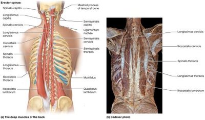
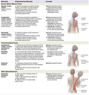
Erector Spinae Muscle Group
Lateral Iliocostalis: Lumborum, thoracis, cervicis.
Middle Longissimus: Thoracis, cervicis, capitis.
Medial Spinalis: Thoracis, cervicis, capitis.
Transversospinal Group
Semispinalis, Multifidus, Rotatores: Extend head, neck, vertebral column.
Quadratus Lumborum: Extends, laterally flexes vertebral column.
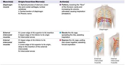
Muscles of Ventilation
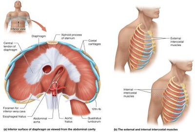
Overview
Diaphragm: Contracts and flattens for inspiration; relaxes for expiration.
External Intercostals: Raise and spread ribs for inspiration.
Internal Intercostals: Depress rib cage for forced expiration.
Sternocleidomastoid and Scalenes: Assist in forced inspiration.
Abdominal Muscles
Overview
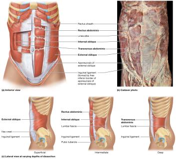
Rectus Abdominis: Flexes trunk, enclosed by rectus sheath, central tendon is linea alba.
External/Internal Obliques: Rotate and flex trunk laterally.
Transversus Abdominis: Compresses abdominal cavity, raises intra-abdominal pressure.
These muscles facilitate urination, defecation, childbirth, and forced expiration.
Muscles of the Pelvic Diaphragm, Urogenital Diaphragm, and Perineum
Pelvic Floor
Levator Ani: Pubococcygeus, Iliococcygeus, Ischiococcygeus; supports pelvic floor, anal canal, genitourinary organs.
Anococcygeal Ligament: Insertion site for pelvic floor muscles.
Perineum
Urogenital Diaphragm: External urethral sphincter (urination control), deep transverse perineal muscle (supports organs).
External Anal Sphincter: Voluntary control of defecation.
Superficial Transverse Perineal, Bulbospongiosus, Ischiocavernosus: Stabilize perineal body, aid erection, expel semen.
Tables: Muscles of the Pelvic Diaphragm, Urogenital Diaphragm, and Perineum
Muscle(s) | Origin/Insertion/Nerve(s) | Action(s) |
|---|---|---|
Levator ani group | O: Interior pelvic bones; I: Coccyx/anococcygeal ligament; N: S3/S4 | Supports pelvic floor, anal canal, genitourinary organs |
Deep transverse perineal | O: Ischiopubic rami; I: Perineal body; N: Pudendal nerve | Supports pelvic organs; fixes central tendon |
External urethral sphincter | N: Pudendal nerve | Compresses urethra (urination control) |
External anal sphincter | O: Coccyx/perineal body; I: Connective tissues around anal canal; N: Pudendal nerve | Closes anal opening |
Bulbospongiosus | O: Perineal body; I: Corpus spongiosum/clitoris; N: Pudendal nerve | Expels semen/urine; assists erection; constricts vagina |
Ischiocavernosus | O: Ischiopubic rami; I: Corpus cavernosum; N: Pudendal nerve | Maintains erection |
Superficial transverse perineal | O: Ischial tuberosity; I: Perineal body; N: Pudendal nerve | Stabilizes perineal body |
Urinary Incontinence and Kegel Exercises
Weak pelvic diaphragm muscles can cause urinary incontinence, especially after childbirth.
Kegel exercises strengthen the levator ani muscle, treating incontinence and preventing pelvic organ prolapse.
Recommended for pregnant women and men with incontinence or sexual dysfunction.
Muscles That Move the Scapula at the Pectoral Girdle
Overview
Serratus Anterior: Protracts scapula, rotates superiorly.
Pectoralis Minor: Protracts and depresses scapula.
Trapezius: Elevates, retracts, depresses, and rotates scapula.
Levator Scapulae: Elevates scapula.
Rhomboid Major/Minor: Retract scapula.
Muscles That Move the Arm at the Shoulder Joint
Overview
Pectoralis Major: Flexes, adducts, and internally rotates arm.
Coracobrachialis: Assists flexion and adduction.
Deltoid: Flexion, abduction, extension of arm.
Latissimus Dorsi: Extends, adducts, internally rotates arm.
Teres Major: Assists latissimus dorsi.
Rotator Cuff: Stabilizes shoulder joint (Teres Minor, Supraspinatus, Infraspinatus, Subscapularis).
Muscles That Move the Forearm and Hand
Forearm at the Elbow Joint
Biceps Brachii: Flexes and supinates forearm.
Brachialis: Agonist of elbow flexion.
Brachioradialis: Assists flexion.
Triceps Brachii: Extends forearm, adducts arm, stabilizes shoulder.
Anconeus: Assists extension.
Hand and Fingers
Flexors: Anterior/medial forearm; flex hand and fingers.
Extensors: Posterior/lateral forearm; extend hand and fingers.
Intrinsic Hand Muscles: Abductor pollicis brevis, flexor pollicis brevis, lumbricals, interossei; move fingers and thumb.
Muscles of the Hip, Thigh, Knee, and Leg
Anterior and Medial Muscles
Iliopsoas: Thigh flexion.
Pectineus: Assists flexion and adduction.
Adductor Group: Magnus, Longus, Brevis; adduct thigh.
Gracilis: Adducts thigh.
Sartorius: Flexion, abduction, lateral rotation of thigh; flexion of leg.
Quadriceps Femoris: Rectus femoris (flexion, extension), vastus lateralis/intermedius/medialis (extension).
Posterior Muscles
Gluteal Group: Maximus (extension, abduction, lateral rotation), Medius/Minimus (abduction, medial rotation).
Piriformis, Obturator Internus/Externus, Gemelli, Quadratus Femoris: Lateral rotation.
Hamstrings: Semitendinosus, Semimembranosus, Biceps Femoris (extension).
Muscles of the Ankle, Foot, and Toes
Foot Movements
Dorsiflexion: Tibialis anterior, extensor digitorum longus.
Inversion: Tibialis anterior.
Eversion: Fibularis longus/brevis.
Plantarflexion: Gastrocnemius, soleus, plantaris, tibialis posterior, fibularis longus/brevis.
Toe Movements
Flexor Hallucis Longus: Flexes hallux.
Flexor Digitorum Longus: Flexes toes 2–5.
Intrinsic Foot Muscles: Support arches, adapt to terrain, skilled movements.
The Big Picture of Muscle Movement
Additional info: All tables have been recreated in HTML format, and only directly relevant images are included. Academic context has been expanded for clarity and completeness. These notes are structured for exam preparation and self-study.
