 Back
BackComprehensive Study Guide for Muscular and Nervous System (Chapters 9–12)
Study Guide - Smart Notes
 Tailored notes based on your materials, expanded with key definitions, examples, and context.
Q1. Classification of Muscles: Based on Actions and Shapes
Background
Topic: Muscle Classification
This question tests your understanding of how muscles are categorized by their actions (e.g., flexors, extensors) and by their shapes (e.g., deltoid, trapezius). Recognizing these categories helps in identifying muscle function and anatomical location.
Key Terms:
Action: The movement a muscle produces (e.g., flexion, extension).
Shape: The physical form of the muscle (e.g., deltoid is triangular, trapezius is trapezoidal).
Step-by-Step Guidance
List the main categories of muscle actions (e.g., flexors, extensors, abductors, adductors).
Identify common muscle shapes (e.g., deltoid, rhomboid, orbicularis).
Match at least one muscle to each action and shape category.
Try solving on your own before revealing the answer!
Q2. Muscles: Location in the Body and Functions
Background
Topic: Major Muscles and Their Functions
This question asks you to identify the location and function of specific muscles, such as the deltoid, biceps, pectoralis, and others. Understanding these helps in mapping muscle anatomy to movement and support.
Key Terms:
Origin: The fixed attachment point of a muscle.
Insertion: The movable attachment point.
Action: The movement produced by the muscle.
Step-by-Step Guidance
For each muscle listed, locate it on a diagram or figure (e.g., deltoid on the shoulder, biceps on the upper arm).
Describe the primary action of each muscle (e.g., deltoid abducts the arm).
Note any groupings (e.g., quadriceps group includes four muscles on the anterior thigh).
Try solving on your own before revealing the answer!
Q3. Muscle Lever Systems
Background
Topic: Biomechanics of Muscle Action
This question focuses on how muscles act as levers in the body, which is essential for understanding movement efficiency and force generation.
Key Terms and Formulas:
Lever: A rigid bar (bone) that moves on a fixed point (fulcrum) when a force is applied.
Fulcrum: The joint around which movement occurs.
Effort: The force applied by muscle contraction.
Load: The resistance moved by the lever.
Step-by-Step Guidance
Identify the three classes of levers (first, second, third) and give an example for each in the human body.
Label the fulcrum, effort, and load for a given muscle action (e.g., biceps curl).
Explain how the arrangement affects force and speed of movement.
Try solving on your own before revealing the answer!
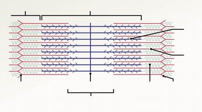
Q4. Sarcomere Microscopic Structure
Background
Topic: Muscle Fiber Anatomy
This question tests your knowledge of the microscopic anatomy of the sarcomere, the functional unit of muscle contraction.
Key Terms:
Sarcomere: The segment between two Z lines in a myofibril.
Actin: Thin filament protein.
Myosin: Thick filament protein.
Z line, M line, A band, I band, H zone: Structural features of the sarcomere.
Step-by-Step Guidance
Label the main structures of the sarcomere on a diagram.
Describe the arrangement of actin and myosin filaments.
Explain the significance of each band and line in muscle contraction.
Try solving on your own before revealing the answer!
Q5. Sliding Filament Theory
Background
Topic: Mechanism of Muscle Contraction
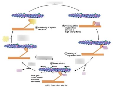
This question is about the process by which muscles contract at the molecular level, specifically how actin and myosin interact.
Key Terms:
Sliding Filament Theory: Explains how muscle fibers contract by the sliding of actin over myosin filaments.
Cross-bridge: The connection formed between actin and myosin heads during contraction.
ATP: Provides energy for the contraction cycle.
Step-by-Step Guidance
Describe the sequence of events in the cross-bridge cycle (attachment, power stroke, detachment, re-cocking).
Explain the role of ATP in each step of the cycle.
Relate the sliding of filaments to the shortening of the sarcomere.
Try solving on your own before revealing the answer!
Q6. Sequence of Muscle Contraction Events
Background
Topic: Muscle Contraction Physiology
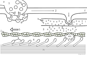
This question asks you to outline the steps from nerve impulse to muscle contraction, including the role of calcium and neurotransmitters.
Key Terms:
Neuromuscular Junction: The synapse between a motor neuron and muscle fiber.
Acetylcholine (ACh): Neurotransmitter that initiates muscle contraction.
Calcium ions (Ca2+): Trigger contraction by binding to troponin.
Step-by-Step Guidance
Describe the release of ACh at the neuromuscular junction.
Explain how ACh binding leads to depolarization of the muscle membrane.
Outline the role of Ca2+ in exposing binding sites on actin.
Try solving on your own before revealing the answer!
Q7. Isometric vs. Isotonic Muscle Contractions
Background
Topic: Types of Muscle Contraction
This question tests your understanding of the differences between isometric (no length change) and isotonic (length changes) contractions.
Key Terms:
Isometric: Muscle tension increases, but length does not change.
Isotonic: Muscle changes length (concentric or eccentric) while moving a load.
Step-by-Step Guidance
Define both types of contractions.
Give an example of each type in the human body.
Explain the physiological significance of each type.
Try solving on your own before revealing the answer!
Q8. Nervous System: General Structure and Functions
Background
Topic: Nervous System Anatomy and Physiology
This question covers the basic organization of the nervous system, including the central and peripheral divisions, and their main functions.
Key Terms:
CNS: Central nervous system (brain and spinal cord).
PNS: Peripheral nervous system (nerves outside CNS).
Neuron: Nerve cell responsible for transmitting signals.
Step-by-Step Guidance
Identify the main divisions of the nervous system.
Describe the primary functions of each division.
Label a diagram of the nervous system with key structures.
Try solving on your own before revealing the answer!
Q9. Synapses, Polarized Neurons, Depolarization, Repolarization, Hyperpolarization
Background
Topic: Neuronal Communication
This question focuses on how neurons transmit signals, including the changes in membrane potential during an action potential.
Key Terms:
Synapse: Junction between two neurons.
Polarization: Resting state of the neuron membrane.
Depolarization: Membrane potential becomes less negative.
Repolarization: Return to resting potential.
Hyperpolarization: Membrane potential becomes more negative than resting.
Step-by-Step Guidance
Describe the sequence of events during an action potential.
Explain the role of sodium and potassium ions in each phase.
Illustrate the changes in membrane potential graphically.
Try solving on your own before revealing the answer!
Q10. Cells of the Nervous System, Saltatory Conduction, Myelin Composition
Background
Topic: Neuroglia and Signal Transmission
This question covers the types of cells in the nervous system, how myelin speeds up conduction, and what myelin is made of.
Key Terms:
Neuroglia: Support cells in the nervous system.
Saltatory Conduction: Rapid transmission of nerve impulses along myelinated axons.
Myelin: Fatty substance that insulates axons.
Step-by-Step Guidance
List the main types of neuroglia (e.g., Schwann cells, oligodendrocytes).
Describe how myelin increases conduction speed.
Explain the composition and function of myelin sheaths.
Try solving on your own before revealing the answer!
Q11. Brain Structures and Functions (Cerebral Cortex, Tracts, Arbor Vitae, Brain Stem, Hypothalamus, etc.)
Background
Topic: Brain Anatomy and Physiology
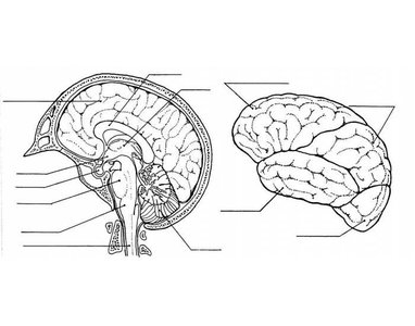
This question asks you to identify and describe the functions of major brain structures, including the cerebral cortex, brain stem, hypothalamus, and more.
Key Terms:
Cerebral Cortex: Outer layer of the brain, responsible for higher functions.
Brain Stem: Controls basic life functions.
Arbor Vitae: White matter in the cerebellum.
Hypothalamus: Regulates homeostasis.
Step-by-Step Guidance
Label the main brain structures on a diagram.
Describe the primary function of each structure.
Relate each structure to its role in the nervous system.
Try solving on your own before revealing the answer!
