 Back
BackComprehensive Study Guide: The Skeletal System (Axial & Appendicular)
Study Guide - Smart Notes
 Tailored notes based on your materials, expanded with key definitions, examples, and context.
The Skeletal System
Overview of the Adult Skeleton
The adult human skeleton consists of 206 bones, divided into two main regions: the axial and appendicular skeleton. Each region contains distinct groups of bones with specialized functions for support, protection, and movement.
Axial Skeleton: 80 bones, including the skull, vertebral column, and thoracic cage.
Appendicular Skeleton: 126 bones, including the pectoral and pelvic girdles, and the limbs.
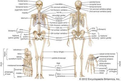
Axial Skeleton
Skull
The skull is composed of 22 bones, divided into cranial and facial bones. Cranial bones protect the brain, while facial bones form the structure of the face and house sensory organs.
Cranial Bones (8): Frontal, parietal (2), temporal (2), occipital, sphenoid, ethmoid.
Facial Bones (14): Maxillae (2), nasal (2), palatine (2), zygomatic (2), lacrimal (2), inferior nasal conchae (2), vomer, mandible.
Key Features: Sutures (immovable joints), foramina (holes for nerves and vessels), and sinuses (air-filled spaces).
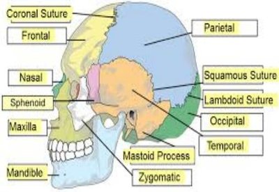
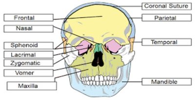
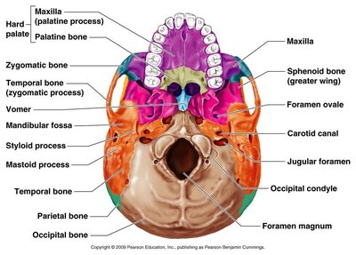
Hyoid Bone
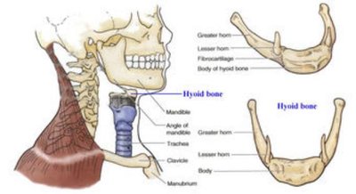
The hyoid bone is a unique, horseshoe-shaped bone located between the chin and larynx. It does not articulate with any other bone and serves as an attachment site for muscles and ligaments involved in swallowing and speech.
Diagnostic Importance: Fracture of the hyoid can indicate strangulation.
Parts: Body, greater horn, lesser horn.
Auditory Ossicles
There are six auditory ossicles (three in each ear): malleus, incus, and stapes. These are the smallest bones in the body and are essential for hearing.
Malleus: Hammer-shaped.
Incus: Anvil-shaped.
Stapes: Stirrup-shaped.
Vertebral Column
The vertebral column consists of 26 bones in adults, organized into five regions. It protects the spinal cord and provides structural support.
Cervical (7): Neck region; includes atlas (C1) and axis (C2).
Thoracic (12): Chest region.
Lumbar (5): Lower back.
Sacrum (1): Five fused vertebrae.
Coccyx (1): Four fused vertebrae (tailbone).
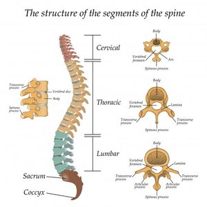
Parts of a Typical Vertebra: Body, pedicle, spinous process, transverse process, vertebral foramen.
Intervertebral Discs: Fibrocartilage between vertebrae for shock absorption.
Disorders of the Vertebral Column
Scoliosis: Abnormal lateral curvature.
Kyphosis: Exaggerated thoracic curvature (hunchback).
Lordosis: Exaggerated lumbar curvature (swayback).
Thoracic Cage
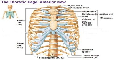
The thoracic cage consists of 24 ribs (12 pairs) and the sternum. It protects vital organs and supports respiration.
True Ribs (1-7): Direct attachment to sternum.
False Ribs (8-10): Indirect attachment via cartilage.
Floating Ribs (11-12): No attachment to sternum.
Sternum: Manubrium, body, xiphoid process.
Sinuses
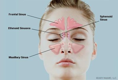
There are four pairs of paranasal sinuses, named for the bones in which they are located. Sinuses lighten the skull and aid in resonance of the voice.
Frontal Sinus
Maxillary Sinus
Ethmoid Sinus
Sphenoid Sinus
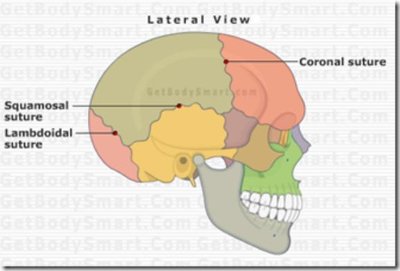
Sutures of the Skull
Sutures are immovable joints between skull bones. In infants, these are fontanels, which later ossify.
Sagittal: Between parietal bones.
Coronal: Between frontal and parietal bones.
Lambdoidal: Between parietal and occipital bones.
Squamous: Between parietal and temporal bones.
Appendicular Skeleton
Pectoral Girdle
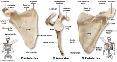
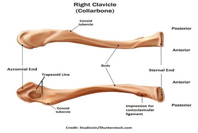
The pectoral girdle consists of the clavicle and scapula, connecting the upper limb to the trunk.
Clavicle: S-shaped bone; sternal end attaches to sternum, acromial end attaches to scapula.
Scapula: Flat, triangular bone; features include spine, acromion, coracoid process, and glenoid cavity.
Pelvic Girdle
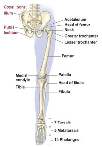
The pelvic girdle transfers the weight of the upper body to the lower limbs. It is formed by the sacrum, coccyx, and hip bones (ilium, ischium, pubis).
Acetabulum: Socket for the head of the femur.
Pubic Symphysis: Cartilaginous joint between pubic bones.
Comparison of Male and Female Pelvis
Feature | Male | Female |
|---|---|---|
Appearance | Massive, heavy | Smoother, less massive |
Acetabulum | Larger | Smaller |
Sacrum | Narrow | Wide |
Coccyx | Less movable, vertical | More movable, tilted |
Pubic Symphysis | Taller | Shorter |
Body | Rounder | Triangular |
Upper Limbs
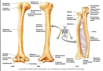
Each upper limb contains 30 bones: humerus (arm), radius and ulna (forearm), carpals (wrist), metacarpals (hand), and phalanges (fingers).
Humerus: Features include head, neck, olecranon fossa, medial epicondyle.
Radius: Lateral bone of forearm; styloid process, head, neck.
Ulna: Medial bone; olecranon process, trochlear notch.
Carpals: 8 wrist bones.
Metacarpals: 5 bones of the palm.
Phalanges: 14 bones; thumb (pollex) has 2, others have 3.
Lower Limbs
Each lower limb contains 30 bones: femur (thigh), tibia and fibula (leg), tarsals (ankle), metatarsals (foot), and phalanges (toes).
Femur: Largest bone; head fits into acetabulum.
Tibia: Medial, larger bone of the leg.
Fibula: Lateral, thinner bone.
Patella: Kneecap.
Tarsals: 7 ankle bones; calcaneus (heel), talus (top of foot).
Metatarsals: 5 bones.
Phalanges: 14 bones; hallux (big toe) has 2, others have 3.
Bone Markings
Types of Bone Markings
Bones have various markings that serve as attachment points for muscles, routes for nerves and blood vessels, and articulation surfaces. These markings are classified as articulations, extensions/projections, depressions, and passages/cavities.
Articulations: Condyle (rounded knob).
Extensions/Projections: Process, tubercle, spine.
Depressions: Alveolus (pit/socket), fossa (shallow basin), fovea (small pit), sulcus (groove).
Passages/Cavities: Canal (tunnel), fissure (slit), foramen (hole), meatus (canal), sinus (air-filled space).
Summary Table: Major Bones of the Skeleton
Region | Main Bones | Key Features |
|---|---|---|
Skull | Frontal, parietal, temporal, occipital, sphenoid, ethmoid, maxilla, mandible | Sutures, foramina, sinuses |
Vertebral Column | Cervical, thoracic, lumbar, sacrum, coccyx | Body, spinous process, vertebral foramen |
Thoracic Cage | Ribs, sternum | True/false/floating ribs, manubrium, xiphoid process |
Pectoral Girdle | Clavicle, scapula | Acromion, coracoid process, glenoid cavity |
Pelvic Girdle | Ilium, ischium, pubis, sacrum, coccyx | Acetabulum, pubic symphysis |
Upper Limb | Humerus, radius, ulna, carpals, metacarpals, phalanges | Olecranon fossa, styloid process |
Lower Limb | Femur, tibia, fibula, tarsals, metatarsals, phalanges | Patella, calcaneus, hallux |
Additional info:
Bone markings and their functions are essential for understanding muscle attachment and joint movement.
Differences in male and female pelvis are important for childbirth and clinical diagnosis.
Sinuses and foramina are critical for the passage of nerves and blood vessels.
