 Back
BackComprehensive Study Notes: Joints (Articulations) in Human Anatomy & Physiology (Ch. 8)
Study Guide - Smart Notes
 Tailored notes based on your materials, expanded with key definitions, examples, and context.
Joints (Articulations) Overview
Introduction to Joints
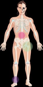
Joints, also known as articulations or arthroses, are the contact points between two or more bones, or between a bone and a tooth. They are essential for skeletal movement and structural stability. The human skeleton contains over 200 bones, most of which form joints with other skeletal elements. Joints may also include cartilage, ligaments, tendons, and muscles.
Key Functions:
Facilitate skeletal mobility
Provide structural stability
Range of Motion vs. Stability: The greater the range of motion, the less stable the joint is.
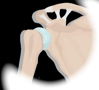
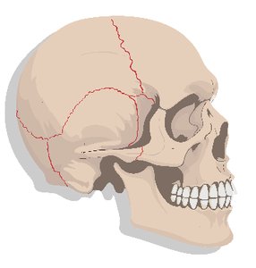
Example: The shoulder joint has the greatest range of motion but is relatively unstable, making it prone to dislocation. In contrast, sutures of the skull are immovable and highly stable.
Classification of Joints
Functional Classification
Joints are classified by the amount of movement they allow:
Synarthroses: Immovable joints (e.g., sutures of the skull)
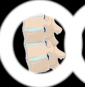
Amphiarthroses: Slightly movable joints (e.g., intervertebral joints)
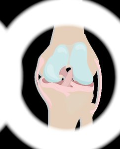
Diarthroses: Freely movable joints (e.g., shoulder, knee)
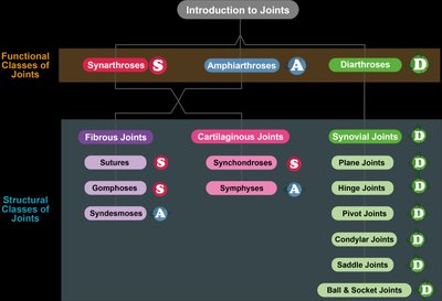
Structural Classification
Joints are also classified based on the material binding the bones:
Fibrous Joints: Bound by collagen fibers of dense connective tissue (always synarthroses or amphiarthroses)
Cartilaginous Joints: Bound by cartilage (always synarthroses or amphiarthroses)
Synovial Joints: Have a synovial cavity and ligaments within an articular capsule (always diarthroses)
Fibrous Joints
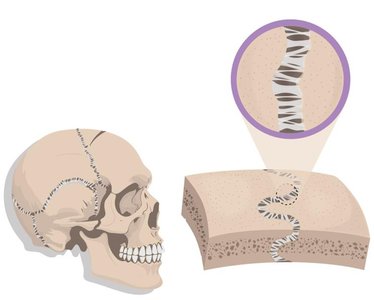
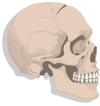
Sutures
Sutures are fibrous joints found only in the skull, composed of dense irregular connective tissue. They provide structural stability and allow no movement. Sutures permit skull expansion during youth, but ossify in adults to form synostoses (bony joints).
Primary Function: Protect the brain
Ossification: Sutures fuse cranial bones into a single bone as development completes
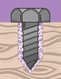
Gomphoses
Gomphoses are fibrous joints that anchor teeth to their bony sockets in the gums. The periodontal ligament, made of dense irregular connective tissue, firmly attaches the tooth to the jawbone. Gomphoses are structurally stable and allow no movement except during chewing or tooth loss in youth.
Periodontal Ligament: "Glues" teeth to their sockets
Development: Ligament deteriorates in youth to allow baby teeth to fall out
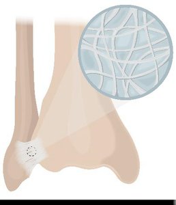
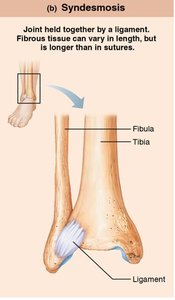
Syndesmoses
Syndesmoses are fibrous joints connecting bones via ligaments made of dense irregular connective tissue. They allow more mobility than sutures or gomphoses, depending on the length of the connecting fibers. Interosseous membranes are broad sheets of connective tissue between certain bones (e.g., radius and ulna, tibia and fibula).
Mobility: Longer fibers = more movement; shorter fibers = less movement
Location: Found in both axial and appendicular skeleton
Cartilaginous Joints
Synchondroses
Synchondroses are cartilaginous joints where bones are bound by hyaline cartilage. They provide structural stability and allow no movement. Some synchondroses ossify over time to become synostoses (bony joints).
Examples: Epiphyseal plates (growth plates) in long bones, costosternal synchondrosis (first rib and sternum)
Ossification: Hyaline cartilage can be replaced by bone
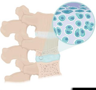
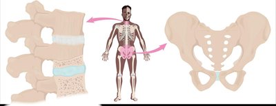
Symphyses
Symphyses are cartilaginous joints located at the body's midline, where bones are bound by fibrocartilage. They are designed for strength and flexibility, allowing a small amount of movement. Fibrocartilage acts as a shock absorber.
Examples: Pubic symphysis, intervertebral joints in the spine
Function: Absorb mechanical stress and provide resilience
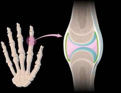
Synovial Joints
General Features
Most joints in the body are synovial joints, which allow dynamic, free movement. They possess several unique characteristics:
Synovial Cavity: Space between bones storing synovial fluid
Synovial Fluid: Viscous liquid serving as a lubricant and shock absorber
Articular Cartilage: Hyaline cartilage covering bone ends, reducing friction
Articular Capsule: Surrounds the joint with two layers:
Inner synovial membrane (produces synovial fluid)
Outer fibrous layer (dense irregular connective tissue)
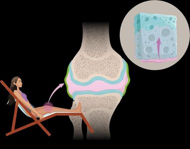
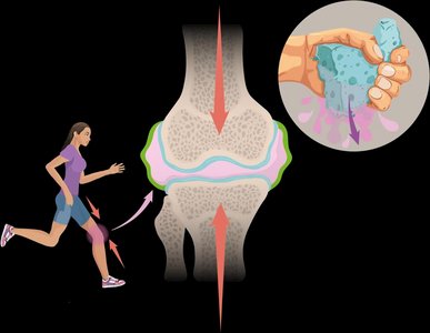
Weeping Lubrication
Weeping lubrication is the process by which synovial fluid is squeezed out of articular cartilage during compression and reabsorbed during decompression. This mechanism lubricates the joint and reduces friction, similar to how a sponge releases and absorbs water.
Articular Cartilage: Stores synovial fluid, reduces stress, and prevents bone ends from direct contact
Additional Features in Synovial Joints
Some synovial joints possess extra structures to enhance function:
Bursae: Synovial fluid-filled sacs that reduce friction between ligaments/tendons and other tissues
Tendon Sheath: Elongated bursa wrapping around a tendon, reducing friction
Fatty Pads: Provide cushioning between fibrous layer and synovial membrane (present in knee and hip joints)
Articular Discs (Menisci): Discs of fibrocartilage or fat dividing the synovial cavity, acting as shock absorbers (present in knee, jaw, etc.)
Summary Table: Joint Classification
Structural Class | Binding Material | Functional Class | Examples |
|---|---|---|---|
Fibrous Joints | Collagen fibers | Synarthroses/Amphiarthroses | Sutures, Gomphoses, Syndesmoses |
Cartilaginous Joints | Cartilage | Synarthroses/Amphiarthroses | Synchondroses, Symphyses |
Synovial Joints | Synovial cavity, ligaments | Diarthroses | Shoulder, knee, finger joints |
Key Terms and Concepts
Articulation: Contact point between bones or bone and tooth
Synarthrosis: Immovable joint
Amphiarthrosis: Slightly movable joint
Diarthrosis: Freely movable joint
Synostosis: Bony joint formed by ossification
Periodontal Ligament: Connective tissue anchoring teeth
Interosseous Membrane: Broad sheet of connective tissue between bones
Synovial Fluid: Lubricant and shock absorber in synovial joints
Bursae: Fluid-filled sacs reducing friction
Meniscus: Fibrocartilage disc acting as shock absorber
Equations and Formulas
While joints are primarily anatomical structures, the following formula relates to joint movement:
Range of Motion (ROM): The degree of movement a joint allows, often measured in degrees.
Example Equation:
Additional info: Academic context was added to clarify the structural and functional classification of joints, the role of connective tissues, and the physiological mechanisms of synovial joints. The summary table and key terms were inferred for completeness and exam preparation.
