 Back
BackComprehensive Study Notes: Nervous System Structure and Function
Study Guide - Smart Notes
 Tailored notes based on your materials, expanded with key definitions, examples, and context.
Nervous System Overview
Functions of the Nervous System
The nervous system is responsible for monitoring the internal and external environment, integrating sensory information, and coordinating voluntary and involuntary responses with other organ systems.
Monitoring: Detects changes inside and outside the body.
Integration: Processes and interprets sensory input.
Coordination: Directs responses by activating muscles or glands.
Organization of the Nervous System
Central Nervous System (CNS) vs. Peripheral Nervous System (PNS)
CNS: Consists of the brain and spinal cord. Damage to the CNS typically does not heal well. Major cell types include astrocytes, ependymal cells, oligodendrocytes, and microglia.
PNS: Communicates with the rest of the body via efferent (motor) and afferent (sensory) pathways. Key cell types are satellite cells and Schwann cells (which are crucial for myelination in the PNS).
Efferent vs. Afferent Pathways
Efferent: Carries motor information from the CNS to muscles and glands.
Afferent: Carries sensory information from the body to the CNS.
Neuron Structure and Function
Anatomy of a Neuron
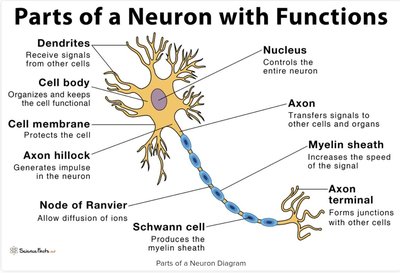
Neurons are the fundamental units of the nervous system, specialized for communication. Each part of a neuron has a specific function:
Dendrites: Receive incoming signals from other cells.
Cell Body (Soma): Contains the nucleus and organelles; essential for cell function.
Axon Hillock: Site where action potentials are initiated.
Axon: Conducts action potentials away from the cell body.
Synaptic Terminals (Axon Terminals): Communicate with other neurons or effectors.
Nucleus: Contains DNA and RNA for protein synthesis.
Nucleolus: Site of RNA synthesis.
Collateral: Branches of the axon ending in synaptic terminals.
Nissl Bodies: Clusters of rough ER and ribosomes, giving gray matter its color.
Structural Types of Neurons
Multipolar: Several dendrites, one axon; most common, especially motor neurons.
Unipolar: Dendrites and axon are continuous; most sensory neurons in the PNS.
Bipolar: One dendrite, one axon; found in special sense organs (e.g., eyes, ears).
Functional Classes of Neurons
Sensory Neurons (Afferent): Transmit sensory information to the CNS.
Motor Neurons (Efferent): Transmit commands from the CNS to effectors.
Interneurons: Connect neurons within the CNS, coordinate motor activity, and process information.
Visceral vs. Somatic Neurons
Visceral Sensory Neurons: Monitor internal environment (organs).
Visceral Motor Neurons: Control involuntary actions (cardiac, smooth muscle, glands).
Somatic Sensory Neurons: Monitor external environment (e.g., proprioception).
Somatic Motor Neurons: Control voluntary movements (skeletal muscle).
Spinal Cord Structure
Meningeal Layers
The meninges are protective membranes surrounding the brain and spinal cord, providing shock absorption, stability, and nutrient supply.
Dura Mater: Outermost, tough layer.
Arachnoid: Middle layer, reduces friction.
Pia Mater: Innermost layer, contains blood vessels.
Ventral and Dorsal Roots
Ventral Roots: Contain axons of motor neurons; join dorsal roots to form spinal nerves.
Dorsal Roots: Contain axons of sensory neurons; dorsal root ganglia house sensory neuron cell bodies.
Gray Horns and White Columns
Gray Horns: Projections of gray matter; posterior horns contain sensory nuclei, anterior horns control skeletal muscle.
White Columns: Myelinated and unmyelinated axons; divided into posterior, lateral, and anterior columns carrying sensory or motor information.
Neuronal Signaling
Depolarization and Hyperpolarization
Resting Membrane Potential: Typically -70 mV; maintained by sodium and potassium gradients.
Depolarization: Interior becomes less negative (more positive), increasing likelihood of action potential.
Hyperpolarization: Interior becomes more negative, decreasing likelihood of action potential.
Action Potentials: Saltatory vs. Continuous Propagation
Continuous Propagation: Occurs in unmyelinated axons; slow, stepwise conduction (~2 mph).
Saltatory Propagation: Occurs in myelinated axons; rapid, jumping conduction between nodes of Ranvier (up to 300 mph).
Divergence vs. Convergence
Divergence: One input spreads to multiple targets (amplification/distribution).
Convergence: Multiple inputs converge on one target (summation/integration).
Brain Structure and Function
Regions of the Cerebrum
Frontal Lobe: Primary motor cortex (voluntary movement), gustatory cortex (taste).
Parietal Lobe: Primary sensory cortex (touch, pain, pressure, temperature).
Occipital Lobe: Visual cortex (vision).
Temporal Lobe: Auditory and olfactory cortex (hearing, smell).
Cerebellum
Coordinates voluntary movements, balance, posture, and motor learning. Acts as a processing center for fine-tuning motor commands.
Association Areas
Interpret incoming sensory and motor information, coordinate responses, and allow recognition of sensory input.
Integrative Centers
Broca's Area: Speech production and language processing (frontal lobe).
Wernicke's Area: Language comprehension (temporal lobe).
Prefrontal Cortex: Executive functions (decision-making, planning).
Diencephalon and Brain Stem
Diencephalon: Includes epithalamus (melatonin production), thalamus (sensory relay), hypothalamus (autonomic and endocrine regulation).
Brain Stem: Controls life-sustaining functions and relays information between brain and body.
Midbrain: Processes visual/auditory info, involuntary motor responses.
Pons: Connects cerebellum to brain stem, motor control.
Medulla Oblongata: Regulates heart rate, breathing, and relays sensory info.
Cranial Nerves
Names and Functions
Olfactory: Smell
Optic: Vision
Oculomotor: Eye movement, lens shape, pupil size
Trochlear: Eye movement
Trigeminal: Sensory from head, motor to chewing muscles
Abducens: Eye movement
Facial: Facial expression, taste, tears, salivation
Vestibulocochlear: Hearing
Glossopharyngeal: Tongue and pharynx movement, taste
Vagus: Sensory/motor to thoracic and abdominal organs
Accessory: Neck and back muscles
Hypoglossal: Tongue movement
Nerve Plexuses
Cervical Plexus: Neck, diaphragm, upper chest, neck, ears
Brachial Plexus: Shoulder girdle, arms
Lumbar Plexus: Pelvic girdle, upper thigh
Sacral Plexus: Hip, leg
Autonomic Nervous System
Sympathetic vs. Parasympathetic
Sympathetic: "Fight or flight"; increases heart rate, respiration, releases energy, inhibits non-essential functions.
Parasympathetic: "Rest and digest"; conserves energy, promotes digestion, slows heart rate.
Sensory Systems
General vs. Special Senses
General Senses: Temperature, pain, touch, pressure, vibration, proprioception.
Special Senses: Olfaction, vision, gustation, hearing, equilibrium.
Types of Sensory Receptors
Nociceptors: Detect pain (temperature extremes, mechanical damage, chemicals).
Thermoreceptors: Detect temperature changes; adapt quickly.
Mechanoreceptors: Detect touch, pressure, vibration, proprioception (includes tactile receptors, baroreceptors, proprioceptors).
Chemoreceptors: Detect chemical changes (pH, CO2 levels); usually subconscious.
Examples of Mechanoreceptors
Tactile Receptors: Fine and crude touch, pressure, vibration (e.g., Merkel's disks, Meissner's corpuscles, Pacinian corpuscles).
Baroreceptors: Monitor pressure in organs; regulate cardiac, respiratory, digestive, urinary functions.
Proprioceptors: Monitor muscle and joint position (muscle spindles, Golgi tendon organs, joint capsule receptors).
Additional info:
Tickle and itch sensations are mediated by the same type of receptor but are perceived differently by the brain.
